
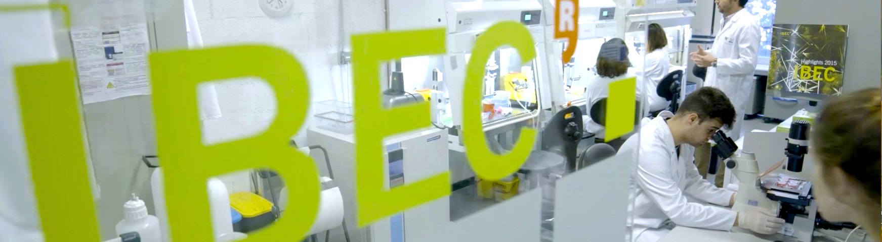
The Institute for Bioengineering of Catalonia (IBEC) is one of the top research institutions named as a Severo Ochoa Research Centre by Ministry of Science and Innovation, which recognizes excellence at the highest international level in terms of research, training, human resources, outreach and technology transfer.
IBEC was established in 2005 by the Ministries of Innovation, Universities and Enterprises and Health of the Generalitat de Catalunya (Autonomous Government of Catalonia), the University of Barcelona (UB) and the Technical University of Catalonia (UPC). Today, IBEC’s relationship with the UB and UPC researchers continues to operate under a framework agreement signed in 2008.
With the aim to train the next generation of experts in bioengineering, IBEC offers in this call 6 PhD fellowships associated to the research projects Generación de Conocimiento 2025, funded by the Spanish Ministry of Science, Innovation and Universities.
LABOUR CONDITIONS
The PhD fellowships offer a 4-year predoctoral contract with the following gross annual salary:
- 24.550,00€ for the 1st year
- 24.550,00€ for the 2nd year
- 24.550,00€ for the 3rd year
- 25.500,00€ for the 4th year
Moreover, 7.000€ is also offered for mobility and training, including the university enrolment fees, during the 4-year period. Indemnities will be paid at the end of the labour contract.
Predoctoral researchers will have to enrol in a university of their choice (mainly University of Barcelona-UB; Polytechnic University of Catalonia-UPC and University Pompeu Fabra-UPF). IBEC doesn’t grant the doctorate degree, instead, it provides the experimental experience you need to complete the PhD. The awarding body of your PhD will be the University at which you are enrolled as a doctoral student.
University enrolment fees will be covered by the fellowship.
PhDs will provide an annual report from the Doctoral School confirming the positive scientific progress related to the PhD thesis carried out during the year. PhDs who finish and defend their PhD thesis before the end of the 4-year period of the fellowship will be able to sign an up to 1-year postdoctoral contract. The aim of this contract is to provide an orientation period to consolidate the knowledge acquired during the PhD thesis and start looking for postdoctoral opportunities, including those through other competitive fellowships. Under no circumstance the total duration of the PhD and postdoc contract cannot exceed 4 years.
The expected initial date is January 1st 2027 (to be confirmed, depending on the publication date of the final resolution of the call Proyectos de Generación de Conocimiento 2025), when a predoctoral contract will be issued , once they have been admitted on a Doctoral Programme.
Other general conditions:
Gross salary provides full social security coverage, which includes health and accident insurance, pension and unemployment benefits. Working conditions at IBEC also include:
- Yearly 23 working days of paid holidays
- 9 leave days for personal matters
- Measures to reconcile work and family life, such as:
- Parental leave (19 weeks)
- Leave for breastfeeding
- Shorter hours for guardianship or leave to care for children and relatives
- Flexible schedule working hours
- Induction programme to facilitate incorporation at IBEC
- Additional support is provided for foreigners to obtain Visa-working permit and to install in Barcelona
IBEC provides Training and PhD discussions specially devoted for PhDs to prepare the thesis and presentation skills. IBEC also provides Seminars with top names in bioengineering and nanomedicine from all over the world in order to offer the opportunity to discuss and network the developments. IBEC also offers different courses to give the opportunity to learn new skills such a leadership communication, time management, and language skills. The institute also holds an annual symposium on a different scientific theme, as well as hosting and organizing several other project-based or general scientific meetings and workshops throughout the year.
As part of the PhD fellowship, PhD’s are encouraged to take up research placements in other centers. Thanks to these stay, young researchers benefit from transnational and multidisciplinary mobility and have the added value of enabling PhD Students to obtain an international PhD, a recognized distinction which significantly improves their chances of a successful career.
All PhDs should commit themselves to participate in outreach and education activities.
In order to enhance and acknowledge the excellence of the training program developed for IBEC’s PhD fellows, we issue a Doctoral Certificate of Excellence funded by the Spanish Ministry of Science and Innovation through the Centro de Excelencia Severo Ochoa award. This certificate is awarded to those fellows who meet the quality requirements of the institute. Additionally, all candidates that received a Doctoral Certificate of Excellence will be eligible for a Doctoral Award. The awardees will receive a prize in an award ceremony at the IBEC Symposium.
Within the Wellbeing Programme, IBEC offers a catalogue of activities on healthy habits, wellbeing and mental health.
IBEC is committed to awareness of diversity and gender equality in science and society. This follows our mission to carry out interdisciplinary research at the highest international quality level which, by crating knowledge, helps to improve health and quality of live, and generate wealth.
REQUIREMENTS
Highly qualified researchers of all nationalities willing to join a stimulating, interdisciplinary research and high-quality scientific environment are welcome to apply.
The following requirements are common to the fellowships available:
- Candidates should be ready to enter an official doctoral programme in January 2027 (under Spanish Law). By this time, they must have obtained a university degree and a master’s degree; or must hold an official university qualification from a country of the European Higher Education Area with a minimum of 300 ECTS of official university studies, of which at least 60 are at master’s level. Candidates who expect to be awarded with such degrees by October 2026 are eligible to apply.
- Candidates must have a strong commitment to scientific research and an excellent academic record.
- Candidates must have good working knowledge of English.
- Candidates must not yet have been awarded a doctoral degree.
- Candidates must not have held a PhD contract exceeding twelve months by the beginning of the fellowship (January 2027).
PhD RESEARCH PROJECTS
We offer 7 PhD Fellowships associated to the following individual research projects:
-
- Group: Cellular and molecular mechanobiology
- PI: Pere Roca-Cusachs Soulere
- Project:(CELLCELLNUC)
- Abstract:
Cells within tissues are constantly submitted to mechanical forces from their microenvironment. These forces regulate cell behaviour in the process of mechanotransduction, which is fundamental for both tissue homeostasis and for its dysregulation in cancer and other diseases. Recent research from our lab and others has shown that forces transmitted from the extracellular matrix (ECM) reach the cell nucleus through integrin-mediated adhesions and the actin cytoskeleton. In response, the nucleus itself acts as a mechanotransducer, regulating transcription, contractility, DNA damage, or chromatin compaction. However, how force is transmitted to the nucleus from cell-cell rather than cell-ECM adhesions, and what regulatory functions this has, is unknown. Our lab has preliminary data suggesting that force transmission through the cell-cell ligand E-cadherin rather than integrins leads to very different mechanotransduction responses in the nucleus. Accordingly, we hypothesize that cell-cell adhesion plays an important role in the regulation of force transmission to the nucleus, that is distinct and potentially opposite to the role of cell-ECM adhesion. Deciphering this role is essential to understand how mechanical signals are integrated at the level of tissues, and the functional implications this has.
Thus, our general aim is to understand how force is transmitted from cell-cell adhesions to the nucleus, and its regulatory implications. We will study this in the context of mammary epithelial cancer cells due to the known importance of mechanical forces in breast cancer, and to the major role of both cell- ECM and cell-cell adhesion in epithelial cell function. In aim 1, we will first establish a toolset for probing mechanical force transmission from cell-cell adhesions to the nucleus. This will include cadherin-coated polyacrylamide hydrogels of tunable stiffness combined with a compatible cell stretch system, additional mechanical techniques, and molecular techniques to interfere with key components of cell-cell adhesions, the cytoskeleton, and the nucleoskeleton. In aim 2, we will unravel how mechanical force transmission from cell-cell adhesions to the nucleus regulates mechanotransduction. To this end, we will perturb the system mechanically and molecularly with the tools of aim 1. Then, we will extend the analysis to systems with progressively increased complexity, first by combining cell-cell and cell-matrix adhesion ligands in the hydrogels, and then by analyzing cell monolayers where cells adhere to the ECM on their basal surface and to other cells on their lateral surfaces. In aim 3, we will evaluate the functional consequences of nuclear mechanotransduction mediated by cell-cell adhesions. We will focus on events known to be triggered by mechanical force application to the nucleus, which includes nuclear translocation of transcription factors, activation of calcium signalling, changes in chromatin methylation, and DNA damage, along with their functional outputs. We will also study specific functions related to breast epithelia and cancer: cell migration, and the epithelial to mesenchymal transition. Once completed, our project will provide important insight into the integration of mechanical forces in epithelial tissues, with potential implications in breast cancer and other pathologies related to mechanical alterations.
-
Reference: Project PID2025-169941NB-I00 funded by MICIU/AEI/10.13039/501100011033 and FEDER, EU.
- Group: Nanoscale bioelectrical characterization
- PIs: Gabriel Gomila Lluch + Annalisa Calò
- Project: (SMARTSPMHISTO)
- Abstract:
Histopathology is the study of the microscopic anatomy of tissues to detect alterations related to diseases to be used in medical diagnostics or to understand the mechanistic of some diseases. Current gold standard histological methods are based on digital optical microscopy combined with sample stage automatic scanners to image the whole histological preparation at the maximum optical resolution. This technology offers detailed information on the tissue structure/composition, through staining, and provides a relatively simple way to store and share the information contained in the histological preparations. One of main challenges of this technology refers to the inter-observer and inter-center interpretation variability. To reduce it, current trends encompass the incorporation of new microscopy modalities to provide additional information to the composition/structure information to enable moving towards a more quantitative digital microscopy, which could be enhanced by artificial intelligence. In the present project we aim at developing a microscopic technique able to add a physical (mechanical and electrical) dimension to histopathology. To this end, we propose to develop a whole slide histological imaging microscopy based on a highly sensitive multiparametric (topography, electrical, mechanical) scanning probe microscope with smart force map scanning modes to provide histopathologists with label-free high-resolution information for improved quantitative diagnostics. To achieve it we will have to push multimodal scanning probe microscopy beyond its current technological limits. On the one hand we will enhance the sensitivity and speed of electrical and mechanical imaging to deal with the challenges posed by tissue section preparations. On the other hand, we will implement smart imaging methods assisted by artificial intelligence, both at the level of the scan area of the microscope and at the level of the scan area of the sample stage, to cope with the traditional slow operation of scanning probe microscopes when applied to very large area samples. Finally, we will develop specific sample preparation methods to enhance simultaneously the different physical properties of the tissues to be measured. We expect to develop a system able to obtain multiparametric (structural, electric and mechanical) physical whole slide images at a resolution below that of optical microscopis and in times that makes this technology viable for practical applications. The microscopic methods will be developed user-friendly and to be fully compatible with conventional optical histology to facilitate its rapid adoption in the medical field. The potential of the developed microscope will be demonstrated with proof-of-concept studies on neurodegenerative and neurovascular diseases, where the addition of the physical dimension in the tissue studies is expected to provide a better mechanistic understanding and a more reliable diagnostic. The results of the present project are expected to constitute a breakthrough in the field of both scanning probe microscopy and medical histology.
- Reference: Project PID2025-170402NB-I00 funded by MICIU/AEI/10.13039/501100011033 and FEDER, EU.
- Group: Nanoprobes and Nanoswitches
- PI: Pau Gorostiza Langa
- Project: (IR-DRUG)
- Abstract:
The molecular and cellular complexity of the brain makes it difficult to understand its workings in health and disease and poses great hurdles to try to restore normal function without creating even bigger disturbances. Epileptic seizures caused by excessive synchronous neuronal activity and altering behavior, motor, and autonomic function are paradigmatic of this problem. They are mediated by several proteins that control neuronal excitability, including glutamate and gamma aminobutyric acid receptors and voltage gated sodium channels. Drugs to adjust the activity of these protein targets have been developed but they produce systemic adverse effects due to the wide expression of these targets, for example in brain regions involved in other functions, or in different organs. Systemic activity generally leads to poorly performing drugs when their benefits outweigh their risks, or to abandoning drug candidates when they do not. This is an intrinsic limitation of conventional pharmacology that is insoluble even with the most selective and potent drugs, and therefore requires new approaches. In principle, adverse effects could be avoided by controlling drug activity on demand at selected locations (e.g. at the seizure onset and brain region). This can be achieved with photoswitchable analogs of the drugs of interest in combination with spatiotemporal patterns of illumination. However, in practice this approach requires drugs activated with safe and tissue-penetrating red and infrared (IR) light, which poses formidable chemical and pharmacological challenges: photochromic molecules activated by IR light are scarce and based on bulky substituents and large conjugated structures that are at odds with the design requirements of small molecule drugs (aqueous solubility, bioavailability, and fine tuning of substituents to achieve high potency and selectivity for the protein target). To crack this problem, systematic optimization of well-established drug scaffolds must be combined with the exploration of novel photochromic groups that offer unprecedented opportunities for noninvasive activation. We have identified a series of red- and IR-photoswitchable drugs for several targets in the central nervous system involved in glutamatergic and gabaergic neurotransmission, nerve impulse propagation (voltage gated sodium channels), and muscarinic and dopaminergic neuromodulation. In the IR-DRUG project, we will expand and refine these compound libraries and their ability to produce non-invasive transcranial activation in vivo, to demonstrate their application in mammalian models of focal epileptic seizures, brain function lost after stroke, attention deficit and hyperactivity disorder, and narcolepsy.
-
Reference: Project PID2025-173990OB-I00 funded by MICIU/AEI/10.13039/501100011033 and FEDER, EU.
- Group: Biosensors for Bioengineering
- PI: Javier Ramón Azcón
- Project: (HEPAGING)
- Abstract:
Aging is a complex biological process characterized by progressive deterioration of multiple organs. Although cellular senescence is recognized as a key factor, the mechanisms by which senescence in one organ initiates and orchestrates systemic aging remain poorly understood. The liver, as a central organ of metabolism and detoxification, plays a critical role in systemic homeostasis. HEPAGING aims to develop and validate sophisticated organ-on-a-chip (OOC) platforms that recapitulate liver-initiated multi-organ aging, providing mechanistic insights into systemic senescence communication and enabling screening for therapeutic interventions. The HEPAGING platform integrates advanced miniaturized biosensing technologies, including localized surface plasmon resonance (LSPR) biosensors with vertical-cavity surface-emitting laser (VCSEL) sources and photodiode detection, transepithelial electrical resistance (TEER) monitoring, to enable real-time, label-free, non-destructive assessment of senescence markers and tissue functional impairment. The modular architecture facilitates seamless exchange among tissue compartments (hepatic, intestinal epithelial, and skeletal muscle), enabling investigation of liver-initiated systemic senescence propagation while maintaining technical flexibility for future expansion into comprehensive multi-organ networks. The project is structured in five integrated work packages: (WP1) Development of aged tissue models through 3D tissue engineering (liver, intestine, muscle) and senescence characterization; (WP2) Integration of the OOC platform with Advanced biosensing technologies (LSPR-VCSEL, TEER); (WP3) Mechanistic studies of inter-organ communication mediated by SASP factors; (WP4) Validation and screening of senolytic and senomorphic compounds; (WP5) Project management, dissemination, and impact. By combining physiologically relevant 3D tissue models with integrated miniaturized biosensing capabilities, HEPAGING will establish an unprecedented experimental platform to investigate the mechanisms by which hepatocellular senescence initiates and orchestrates systemic aging through inter-organ communication mediated by the senescence-associated secretory phenotype (SASP). The project will enable identification of tissue-specific intervention targets and biomarkers to prevent or reverse multi-organ aging phenotypes at their systemic source, with direct applications in the development of anti-aging therapies and personalized medicine based on senescence biomarkers.
- Reference: Project PID2025-175576OB-I00 funded by MICIU/AEI/10.13039/501100011033 and FEDER, EU
- Group: Biosensors for Bioengineering
- PI: Juan Manuel Fernández Costa
- Project: (MY2DREAM)
- Abstract:
Myotonic dystrophy type 2 (DM2) is an autosomal dominant genetic disorder caused by an unstable CCTG repeat expansion in the CNBP gene, leading to nuclear RNA foci that sequester splicing regulators like MBNL1 and RBFOX proteins, thereby disrupting alternative splicing of numerous transcripts critical for muscle function and tissue homeostasis. This results in progressive proximal muscle weakness, exercise-induced myalgia, myotonia, early hip girdle involvement, cardiac conduction abnormalities, cataracts, insulin resistance, and milder cognitive effects compared to myotonic dystrophy type 1 (DM1), which arises from CTG expansions in the DMPK gene and features more severe distal-predominant weakness, congenital forms, facial and neck muscle involvement, hypersomnia, and greater respiratory compromise. While DM1 has advanced further in therapeutic development with several antisense oligonucleotides and small molecules reaching clinical trialsDM2 lacks equivalent preclinical platforms, hampering drug repurposing efforts despite molecular overlaps in RNA toxicity mechanisms. The MY2DREAM project addresses this disparity by engineering innovative, patient-derived 3D skeletal muscle tissues to accelerate repurposed drug discovery for DM2. Our central hypothesis asserts that these advanced in vitro human models will faithfully recapitulate DM2-specific pathogenesisincluding molecular, structural, and functional phenotypes as splicing defects, fiber type changes, muscle weakness and delayed relaxation (myotonia), far surpassing the limitations of 2D cultures or non-human models.
MY2DREAM is structured around three synergistic specific aims: (1) Generate and comprehensively validate functional 3D DM2 muscle tissues from patient myoblasts via structural, molecular, and functional assays to confirm disease fidelity across patient genotypes and disease severities, (2) Execute high-throughput screening (HTS) of a curated 5,272-compound repurposing library from MedChemExpress (prioritizing FDA-approved drugs and phase I trial completers) using automated liquid handling robotics, high-throughput imaging, and multiparametric analytics, and (3) Perform in-depth validation of the HTS hits alongside anti-DM1 molecules currently in clinical trials through dose-response studies in the 3D tissues, evaluating functional outcomes, molecular endpoints, and structural improvements, thereby de-risking candidates for DM2 with cost savings over animal or iPSC-derived alternatives.
By leveraging DM2 patient heterogeneity for stratified modeling, MY2DREAM not only deepens mechanistic insights into proximal myopathy and systemic features but also enables personalized therapeutic strategies, bridging the translational gap between DM1 successes and DM2 unmet needs. Ultimately, this project promises to expedite clinical candidates, reduce development timelines through human-relevant phenotyping, and catalyze effective interventions for DM2 patients worldwide.
-
Reference: Project PID2024-162521OB-I00 funded by MICIU/AEI and FEDER, EU.
- Group: Nanobioengineering
- PIs: Anna Lagunas Targarona + Mònica Mir Llorente
- Project: (GBBRAIN-T)
- Abstract:
Gliomas are a group of brain tumors derived from glial cells that range from low-grade forms to highly aggressive types such as glioblastoma (GBM). GBM is the most common malignant primary brain tumor in adults and is associated with severe clinical, social, and personal burdens due to rapid progression, neurocognitive decline, disability, and heavy caregiving demands. Despite maximal standard therapy, including surgery and chemoradiotherapy, survival remains poor, with median survival around one year and scarce long-term survival. Resistance, invasiveness, and pronounced heterogeneity limit current treatments.
Immunotherapies, particularly chimeric antigen receptor (CAR)-T cells, have transformed hematologic cancer care. CAR-T cells are engineered to recognize tumor-associated antigens and kill malignant cells, however their application in solid tumors is hindered by limited antigen specificity, heterogeneous expression, poor penetration into dense tissues, and suppressive tumor microenvironments. In brain tumors, the BBB, complex neuroanatomy, and risks of neurotoxicity add further challenges, as cytokine-mediated endothelial activation can impair barrier integrity and trigger neuroinflammation.
To overcome these obstacles, preclinical testing requires advanced human-relevant models that reproduce the neurovascular environment of GBM. Traditional 2D cultures and animal models fail to capture the architectural, cellular, and microenvironmental complexity of human brain tissue. A robust in vitro model must incorporate a functional BBB and the full neurovascular unit, since vasculature governs tumor invasion, metabolic support, and drug accessibility. Reproducing GBM-BBB interactions enables realistic evaluation of vessel remodeling, barrier disruption, glial and pericyte reactivity, and therapeutic permeability.
Organ-on-a-chip technologies provide microphysiological platforms capable of recreating human tissue complexity with translational relevance. Building on the broad expertise in BBB-oC and neurovascular models from our group at IBEC, this project aims to develop a fully human GBM-BBB-oC System integrating neurons, glia, endothelial cells, and tumor cells. The platform will incorporate dual real-time monitoring: TEER for continuous assessment of BBB integrity and MEA-based electrophysiology to track neuronal network functions. To address limitations of planar MEAs in 3D cultures, the project will explore next-generation 3D MEA designs that improve signal capture and depth-resolved recordings.
In collaboration with the clinics, CAR-T therapies will be evaluated on-chip to study T-cell infiltration, cytotoxicity against GBM cells, interactions with neurovascular components, and potential adverse effects such as vascular dysfunction or neurotoxicity. The system will be designed for scalability, automation, and compatibility with industrial workflows to enhance translational impact. Supported by strong prior experience integrating BBB and neuronal co-cultures with TEER and MEA readouts, the project proposes an innovative human -based neurovascular GBM-BBB-oC platform for mechanistic studies and immunotherapy testing. This approach has the potential to accelerate preclinical discovery, improve prediction of therapeutic efficacy and safety, and ultimately contribute to better outcomes for patients facing one of the deadliest brain cancers.
- Reference: Project PID2024-161645OB-I00 funded by MICIU/AEI/10.13039/501100011033 and FEDER, EU.
SELECTION PROCEDURE
IBEC holds the HR Excellence in Research Award in recognition of our ongoing commitment. The recognition by the European Commission has been renewed several times, last one in February 2022.
Our Recruitment and Selection Policy is based on the OTM Strategy (Open, Transparent and Merit-based recruitment) http://www.ibecbarcelona.eu/jobs/ and accept applications without distinction on any grounds. Candidates with disabilities are strongly encouraged to apply. Our commitment to OTM-R principles can also be found in our Gender and Diversity plan. In line with the principles defined in the OTM-R procedure, selection processes are governed by the following principles:
- Transparency throughout the whole process
- Equal opportunities in the selection and hiring of personnel
- Non-discrimination on grounds of sex, age, ethnic, national or social origin, religion, sexual orientation, language, disability, political opinions or social and economic condition
- Merit based evaluation
- Confidentiality as the cornerstone of the selection process
- Principle of public dissemination of the selection processes, which must also be internationally comparable
Applications will be reviewed by a selection committee led by the corresponding IBEC Principal Investigator of the Research Group.
| Criteria | Score |
|---|---|
| 1. Academic and professional career of the candidate | 0–50 |
| a) Scientific and technological contributions | 0–45 |
| b) Mobility and internationalization | 0–5 |
| 2. Adequacy of the candidate to the research activities to develop | 0–50 |
| TOTAL SCORE | 100 |
CALENDAR AND USEFUL DATES
-
September 21st, 2026: Launch of the call.
-
October 20th, 2026: Deadline for submission of applications.
-
October 21 st – November 20, 2026: Evaluation of the candidates: CVs and online interviews.
-
December 1st, 2026: Communication of the results: the acceptance letters will be sent to the shortlisted candidates. Applicants who have not been successful but have received a positive evaluation will be put on a waiting list for future positions in case a final candidate withdraw the offer.
-
As of January 1st, 2027: Start of the predoctoral contract (to be confirmed, based on the resolution of the call Proyectos de Generación de Conocimiento 2026 by the Spanish Ministry of Science, Innovation and Universities).
CONTACT
If you have any further questions about PhD fellowships, or if there are particular issues you’d like to discuss regarding your application, please contact phd@ibecbarcelona.eu
Institute for Bioengineering of Catalonia (IBEC) www.ibecbarcelona.eu/phd
