
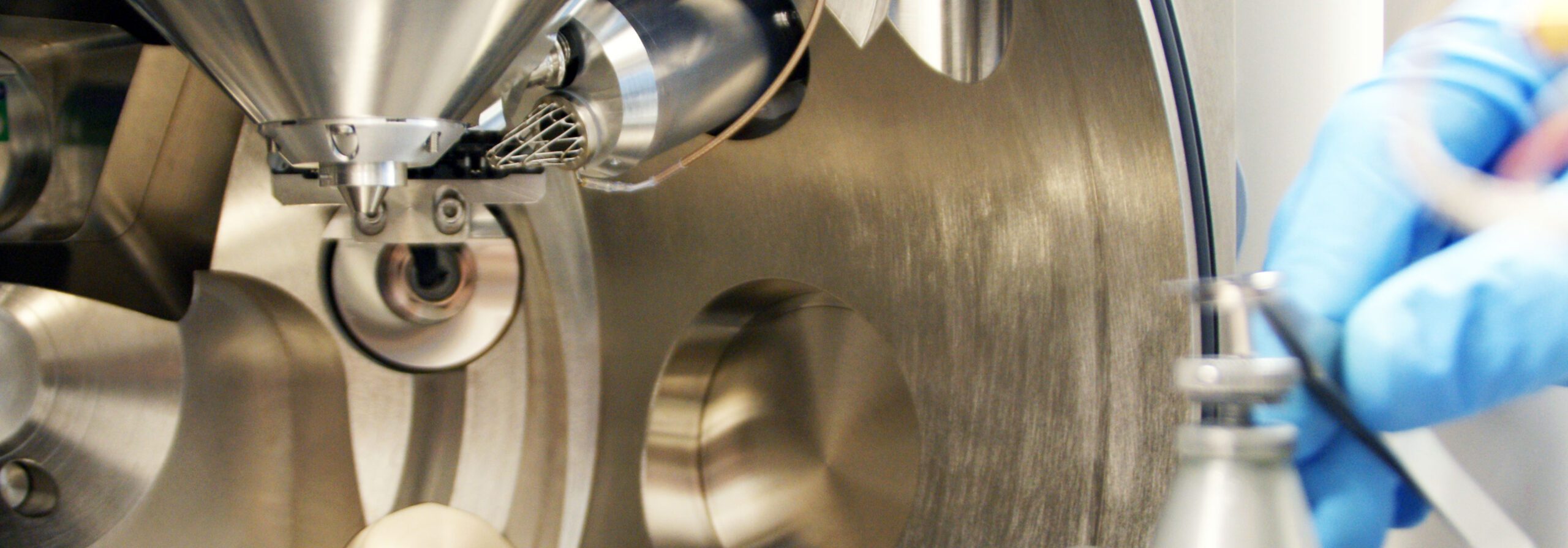
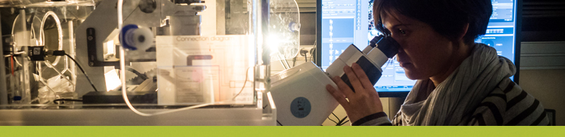
The Microscopy Characterization facility is composed of a variety of microscopy equipment, useful for very different applications in the biomedical field.
With these techniques IBEC researchers can acquire images and analyse structures, from single molecules all the way to the nanoscale of living cells.
We have a combined service located at different spaces and managed separately. Therefore, we offer microscopes managed centrally by the Core Facilities Unit such as the SEM and Confocal microscopes, which are open to other public and private institutions. There are also other instruments managed by IBEC groups that open 30% of their usage time to other IBEC researchers.
For more information please send an email to microscopy@ibecbarcelona.eu.
To make equipment reservations, click the button (only for registered users): 
Equipment
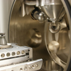
Managed by Core Facilities
Click on an item for more information.
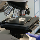
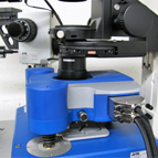
Managed by IBEC groups
Click on an item for more information.
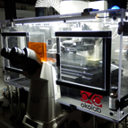
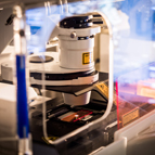
Reconstruction Super
Resolution Microscope
Services
Training as self-user on the available microscopy tools (only for IBEC users).
Custom microscopy services (quotation required).
- SEM morphological and topographical characterization.
- Preparation of SEM samples:
– Gold coating service for high resolution inspection of insulating samples.
– Chemical fixation of biological samples. - Confocal and fluorescence imaging.
- Topographical (SEM) and fluorescence (Confocal) combined studies.
Price lists
 External rates (English)
Visits
If you would like to visit IBEC’s Microscopy Characterization Facility, please fill out the corresponding application form with the information requested. Then send it to microscopy@ibecbarcelona.eu.
Your request will be reviewed, and we will contact you to confirm the day and time of your visit.
Visit request form (English)
How can I become a user?
To become a Microscopy Characterization user, please follow the steps below.
- 1. Read the ‘information and regulations for users’ document:
Terms and conditions of use (internal users)
Terms and conditions of use (external users) - 2. Fill in the new user application forms:
a. Customer registration form: to be completed by the person who assumes the costs resulting from the provision of services carried out in Microscopy Characterization.
Customer Registration Request
b. User linked to a customer registration form: to be completed by the person that will directly use Microscopy Characterization services. This person must be linked to a customer that will assume the costs resulting from the services provided.
User linked to customer registration form (internal users)
User linked to customer registration form (external users)
- 3. Send the completed application forms to microscopy@ibecbarcelona.eu
Our external clients
| Organisation / Group(s) | |
|---|---|
 |
GP-Pharm S.A. Drug Delivery Research |
| Infinitec Activos S.L. |
|
 |
Institut Químic de Sarrià Bioenginyeria |
 |
Universidad del Pais Vasco Tecnología Farmacéutica |
| Institut de Química Avançada de Catalunya (IQAC-CSIC) Nanotecnologia Química y Biomolecular |
|
| Fundació Institut Català Nanociència i Nanotecnologia (ICN2) Advanced Electronic Materials & Devices |
|
| Universitat de Barcelona Física Aplicada y Òptica |
Users' publications
Why acknowledge Microscopy Characterization and its staff in your publications?
- To demonstrate the value of IBEC’s common facility and staff to the scientific community
- To secure funding and new investments in future equipment and better facilities
- To make research better, and keep improving
For every publication accepted by a journal that properly refers to the use of IBEC’s Microscopy Characterization, authors will receive a free hour of instrument use (excluding consumables). Simply inform us about your publication to receive your free time. In addition, we’ll also display your publication on our web page.
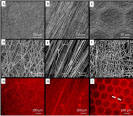 |
Nedjari S, Awaja F, Altankov G. (2017). Three Dimensional Honeycomb Patterned Fibrinogen Based Nanofibers Induce Substantial Osteogenic Response of Mesenchymal Stem Cells Sci Rep. 7(1):15947 |
 |
H A Moreno, S Hussain, R Amade and E Bertran (2014). Growth and functionalization of CNTs on stainless steel electrodes for supercapacitor applications. Materials Research Express, Volume 1, Number 3 |
 |
Ziqiu Tong, Oscar Seira, Cristina Casas, Diego Reginensi, Antoni Homs-Corbera, Josep Samitier and J. Antonio Del Rio (2015). Engineering a functional neuro-muscular junction model in a chip RSC Adv. |