A new study recently published in the journal Scientific Reports, and led by the Institute for Bioengineering of Catalonia (IBEC) in collaboration with MIT, sheds new information on the role of proprioceptive sensory neurons in ALS disease.
Amyotrophic lateral sclerosis (ALS) is a deadly neuromuscular disease that causes approximately 30,000 deaths annually worldwide. Patients usually live 2 to 5 years after diagnosis, and there is currently no cure or effective treatment.
The main feature of ALS is the progressive degeneration of motor neurons in the brain and body, which leads to the gradual loss of voluntary motor function. Although sensory neurons that receive signals from the environment (for example, light and sound) tend not to be affected by ALS, studies carried out in mouse models have revealed that degeneration does occur in proprioceptive sensory neurons, which are responsible for perceiving the position, movement and tension of muscles. joints and tendons of the body itself.
Proprioceptive sensory neurons are central to the sense of proprioception, which allows people to perceive the location and movement of their limbs without needing to see them. Although these neurons have been little studied in relation to ALS, it is believed that their degeneration may contribute to the impairment of motor function in this disease.
A recent study, published in the journal Scientific Reports and led by the Institute of Bioengineering of Catalonia (IBEC), provides evidence on the role of proprioceptive sensory neurons in ALS disease, suggesting that these cells may play an active role in the progression of the disease, thus its study may be key to understanding the pathology and developing more effective treatments.
This study has been led by Josep Samitier, principal investigator of the Nanobioengineering group at IBEC and professor at the University of Barcelona, and Dr. Maider Badiola-Mateos, IBEC researcher and currently affiliate to the Scuola Superiore Sant’Anna in Italy, as lead author. In addition, it had the collaboration of researchers from the Massachusetts Institute of Technology (MIT).
Unraveling the role of proprioceptive sensory neurons
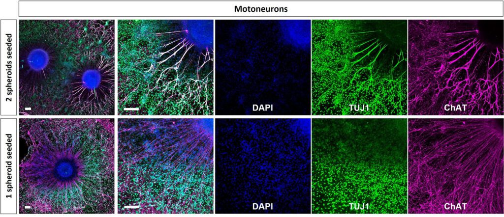
The research compared in vitro models of healthy neuromuscular cells affected by ALS. Human stem cells, both healthy and ALS-affected, were used and differentiated into proprioceptive sensory neurons. Then, their connection to skeletal muscle cells was established using a microfluidic device to observe how these interactions changed. In this way, it was possible to observe for the first time in the laboratory the connection of these human neurons with muscle fibers, including synaptic buttons.

Genetic analysis revealed several differences between healthy and ALS-affected neuromuscular cell models, including lower expression of the ETV1 gene in proprioceptive sensory neurons in ALS. Previous studies had shown that this gene is essential for the formation of functional connections between proprioceptive sensory neurons and motor neurons. Therefore, a reduction in the expression of the ETV1 gene may be relevant for the degradation of the connection of proprioceptive sensory neurons observed in ALS pathology.
Proprioceptive sensory neurons are responsible for muscle stretching myotatic reflexes and therefore this study may explain why muscles lacking this reflex (such as those in the eyes, anus or urethra) are the last to degenerate in ALS patients. In addition, this work suggests that the expression of the ETV1 gene could be induced by substances secreted by target cells. Future research that can mimic a cellular environment capable of inducing proper gene expression of this gene may be crucial to understanding the role of each cell in the development of ALS and designing effective treatments against the disease.
Referenced article:
Badiola-Mateos, M., Osaki, T., Kamm, R.D. & Samitier, J. In vitro modelling of human proprioceptive sensory neurons in the neuromuscular system. Sci Rep 12, 21318 (2022). https://doi.org/10.1038/s41598-022-23565-3


