
ABOUT
The main goal of the Nanoscale Bioelectrical Characterization group is to develop a multiscale and multimodal (electrical, mechanical) approach to Bioelectricity, covering from the nano- to the microscale. To this end the group combines methods and techniques from Scanning Probe Microscopy, Artificial Intelligence and Organic Bioelectronics. The main objective is to contribute to develop new label-free characterization tools for Life Sciences, new nanomedical diagnosis approaches and new electronic biosensors.
Autonomous multimodal scanning probe microscopes for Life Sciences
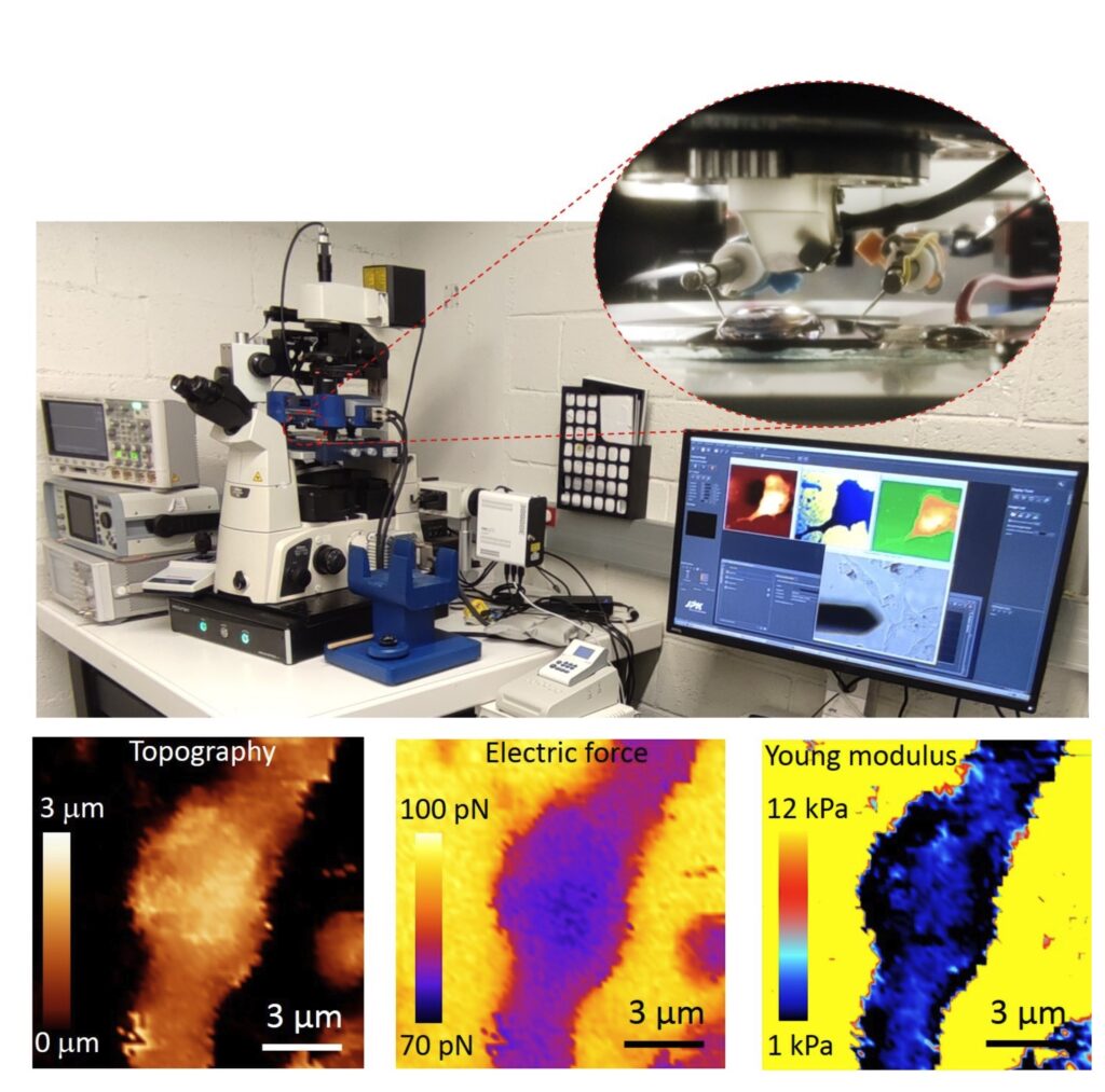
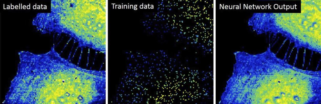
At present the group focuses in the development of an Autonomous Multimodal Functional Scanning Probe Microscope assisted by Artificial Intelligence for Life Sciences and Medical applications. The objective is to map the structural, electrical and mechanical properties at the nanoscale of cells, bacteria, drug nanocarriers and organic Bioelectronic devices with minimal intervention of the operator and at high throughput.
The objective is to obtain in an autonomous way fast functional electric and mechanical nanoscale maps of Life Science samples and Organic Electronics devices in physiological conditions with minimal intervention of the operator and at high throughput.
Initial results obtained by the group include the upgrade of the Scanning Dielectric Microscope to enable its operation in physiological buffers for living cell imaging, the development of a supervised machine learning algorithm to process Scanning Dielectric Microscopy data and provide almost instantaneously local dielectric constant maps of both eukaryotic and prokaryotic cells, and the implementation of a workflow for Scanning Dielectric Microscopy for high throughput and automatic nanoscale multimodal (electrical and mechanical) characterization.
High throughput multimodal characterization of drug nanocarriers
The development of novel drug nanocarriers require an exhaustive multiparametric characterization, which includes its morphology and structure, net charge, particle size distribution or phase transition temperature. These characteristics are obtained usually from different techniques. We target to obtain simultaneously and at high throughput multiparametric information on drug nanocarriers by using a single instrument, namely, the autonomous multimodal in liquid Scanning Dielectric Microscope. We aim at obtaining information on the size, sphericity, membrane wall thickness, lamellarity, Young’s modulus, stiffness, surface charge and membrane specific capacitance of drug nanocarriers, such as liposomes, polymeric nanoparticles or lipid nanoparticles.
Interrelation of mechanical and electrical processes in living neurons
Mechanical and electrical processes in cells and tissues can sometimes appear interrelated, as for instance, in the action potential propagation in neurons, which provokes the electrical polarization of the cell membrane and, at the same time, a change in neuron’s membrane tension. Similarly, the restructuring of the cytoskeleton of neurons, as occurring in the Alzheimer disease, can induce a change in cellular stiffness and, consequently, an improper neuron firing. We aim at investigating this interrelation by means of the multimodal in liquid Scanning Dielectric Microscope applied to living neurons.
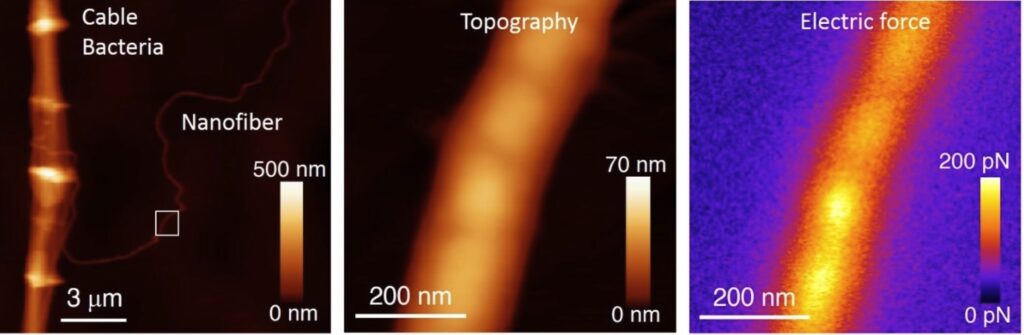
Unravelling the electrical conduction properties of cable bacteria
Long-range electron conduction in cable bacteria filaments presents unusual characteristics in the biological world, exceeding by more than 6 orders of magnitude the conductivity of the best conducting protein nanowires. Electric conduction takes place through Niquel rich protein nanofibers located in the bacteria periplasm, but still many aspects of the electronic conduction in cable bacteria remain unknown. We aim at providing new insights on the conducting properties of cable bacteria by using the unique capabilities and versatility of the Scanning Dielectric Microscope.
Novel nanoscale physical phenotyping of cancer cells
The whole process of cancer aggression, from local growth to extravasation into blood vessels, migration, seeding into different organs and formation of metastases involves physical changes (mechanical and electrical) and their interplay with protein expression and genetic transformations. We aim at developing a high throughput nanoscale multimodal physical phenotyping method for cancer cells based on the Scanning Dielectric Microscope. Our ling term objective is to provide additional diagnostics tools to medical doctors for evaluating cancer progression and aggression.
Structure-function relationships for materials in Organic Bioelectronics
Organic semiconductor materials have emerged as key materials in the development of platforms (e.g. electrolyte gated transistors) for transducing and amplifying biological and biochemical signals. This fact makes them an integral part of diverse biosensing and bioelectronic devices able to sense even single molecules or to record bioelectric potentials from excitable cells. The fundamental understanding of the nanoscale electronic and ionic transport governing the operation of these materials and devices remains, however, poorly understood. We aim at providing new insights into the structure-function relationship of organic materials used in Bioelectronics with the unique capabilities of the multimodal in operando in-liquid Scanning Dielectric Microscope.
STAFF
Staff members:
 ibecbarcelona.eu
Former members:
Harishankar Balakrishnan | PhD Student
Now: Post-doc, University of Munich (Germany)
Ignacio Casuso | PhD Student
Now: Staff Scientist, INSERM (France)
Maria Chiara Biagi | PhD Student
Now: In-vivo Image Analysis Scientist, AstraZenca (Spain)
Marti Checa | PhD Student
Now: R&D Staff scientist, Oak Ridge National Laboratory (USA)
Martin Edwards | Postdoc
Now: Assistant Professor, University of Arkansas (USA)
Daniel Esteban Ferrer | PhD Student
Now: CEO, ViR S.L. (Spain)
Laura Fumagalli | Senior Researcher
Now: Reader, University of Manchester (UK)
Georg Gramse | PhD Student
Now: Group Leader, Johannes Kepler University of Linz (Austria)
Larisa Huetter | PhD Student
Now: IT consultant, Rewion (Germany)
Adrica Kyndiah | Postdoc
Now: Senior Scientist, Instituto Italiano di Tecnologia (Italy)
Helena Lozano | PhD Student
Now: Project Manager, CSIC (Spain)
Martina di Muzzio | PhD Student
Now: Engineer PMQ, Roche (Spain)
Jordi Otero | Postdoc
Now: Lecturer, Universitat de Barcelona (Spain)
Shubham Tanwar | PhD Student
Now: Post-doc, Italian Institute of Technology (Italy)
Romen Trujillo | PhD Student
Now: Associate Professor, Universitat de Barcelona (Spain)
Marc Van der Hofstadt | PhD Student
Now: Post-doc, CNRS (France)
PROJECTS
| INTERNATIONAL PROJECTS | FINANCER | PI |
|---|---|---|
| PRINGLE · Protein Based Next Generation Electronics (2022-2026) | European Commission, PathFinder Open | Gabriel Gomila |
| SPM4.0 · Autonomous Scanning Probe Microscopy for Life Sciences and Medicine powered by Artificial Intelligence | European Commission , MSCA-DN 2023 | Gabriel Gomila |
| NATIONAL PROJECTS | FINANCER | PI |
|---|---|---|
| ICREA Academia Award (2023-2027) | Catalan Institution for Research and Advanced Studies (ICREA) / Generalitat de Catalunya | Gabriel Gomila |
| Microscopio de fuerzas de barrido multiparamétrico autónomo y de alto rendimiento para aplicaciones en ciencias de la vida y medicina (BIOMEDSPM4.0) | MICIU/AEI and FEDER, UE | Gabriel Gomila |
| SGR-Grups de recerca consolidats (SGR-Cat 2021)_GRC | AGAUR / SGR | Gabriel Gomila |
| FINISHED PROJECTS | FINANCER | PI |
|---|---|---|
| SGR Grups de recerca consolidats (2017-2020) | AGAUR / SGR | Gabriel Gomila |
| SPM2.0 · Scanning probe microscopies for nanoscale fast, tomographic and composition imaging (2017-2020) | Marie Curie Skłodowska European Training Network (MSCA-ITN-ETN) | Gabriel Gomila (Project Coordinator) |
| NANOMICROWAVE · Microwave Nanotechnology for Semiconductor and Life Sciences (2013-2016) | MARIE CURIE – ITN | Gabriel Gomila |
| V-SMMART Nano · Volumetric Scanning Microwave Microscopy Analytical and Research Tool for Nanotechnology (2012-2016) | NMP – SME | Gabriel Gomila |
| AFM4NanoMed&Bio · European network on applications of Atomic Force Microscopy to Nanomedicine and Life Sciences | EU COST Action TD1002 | Gabriel Gomila (Management Committee Substitute Member) |
| BIOWIRESENSE · Plataforma universal para la detección de biomarcadores basada en nanocables bacterianos conductores (2017-2019) | MINECO, Explora Ciencia | Gabriel Gomila |
| NANOELECTOMOGRAPHY· Electrical nanotomography based on scanning probe microscopy for nanomaterials and biological samples (2014-2016) | MINECO (TEC2013-48344-C2-1-P) | Gabriel Gomila |
| NANOELECTROPHYS · Scanning Electric Force Microscope for Electrophysological Recordings at the Nanoscale (2016-2019) | MINECO (TEC2016-79156-P) | Gabriel Gomila |
| ICREA Academia Award (2015-2019) | Catalan Institution for Research and Advanced Studies (ICREA) / Generalitat de Catalunya | Gabriel Gomila |
| BORGES · Biosensing with ORGanic ElectronicS (2019-2022) | Marie Curie Skłodowska European Training Network (MSCA-ITN-ETN) | Gabriel Gomila |
| BIGDATASPM · Métodos de datos masivos aplicados a la Microscopía de Sonda de Barrido para estudios eléctricos funcionales en ciencias de la vida (2020-2023) | MINECO, Generación Conocimiento: Proyectos I+D | Gabriel Gomila |
| Correlative Electrical and Mechanical Scanning Probe Microscopy for Life Science Application | Beatriu de Pinós 2019/ AGAUR | Aurora Dols |
PUBLICATIONS
Check for more detailed information on the outputs of the Group at IBEC CRIS portal.
Publications list:
EQUIPMENT
- Cypher Atomic Force Microscope (Asylum Research)
- Nanowizard 4 Bio-Atomic Force Microscope (JPK)
- Cervantes Atomic Force Microscope (Nanotec Electronica)
- Easy Scan 2 Atomic Force Microscope (Nanosurf)
- AxioImager A1m Reflection Optical Microscope (Zeiss) equipped with a AxioCam ERc5s (Zeiss)
- CompactStat portable electrochemical interface and impedance analyzer (Ivium Technologies)
- Palmsens 4, 8 channel Potentiostat (Palmens)
- 2 eLockIn204 4-phase Lock-In amplifiers (Anfatec)
- Keithley 6430 sub-femtoAmp remote sourcemeter
- Keysight B2912A precision Source/Measure Unit, 2 channels
- Keysight N9310A RF Signal Generator 9 kHz to 3.0 GHz
- Computation Workstation Intel Xeon, NVIDIA RTXA5000
COLLABORATIONS
- Dr. Filip Meysman
University of Antwerp, Belgium - Dra. Adrica Kyndiah
Italian Institute of Technology, Italy - Dr. Martí Checa
Oak Ridge National Laboratory, USA - Dr. Jordi Borrell
University of Barcelona, Spain - Dra. Marta Mas-Torrents
Institut de Ciències de Materials de Barcelona, Spain - Dr. Eduard Torrents
Institut de Bioenginyeria de Catalunya, Spain - Dr. Jose Antonio del Rio
Institut de Bioenginyeria de Catalunya, Spain
NEWS
Un nou estudi sobre la síntesi d’ADN en bacteris impulsa la recerca de la pròxima generació d’antibiòtics
Un estudi dirigit per l’Institut de Bioenginyeria de Catalunya (IBEC) i l’Institut de Biologia Molecular de Barcelona (IBMB) ofereix la imatge més detallada fins a la data de l’NrdR, el regulador principal dels ribonucleòtids (RNR) en els bacteris. Els investigadors van obtenir les primeres imatges detallades de l’estructura completa de la proteïna l’NrdR i van demostrar com els canvis en la forma i l’associació d’aquesta proteïna afecten la manera en què controla processos clau dins de la cèl·lula. Les troballes, publicades recentment a la revista International Journal of Biological Macromolecules, amplien la nostra comprensió de com els bacteris regulen la producció dels components moleculars de l’ADN, un aspecte crucial tant per a la microbiologia fonamental com per al desenvolupament de noves estratègies antimicrobianes.
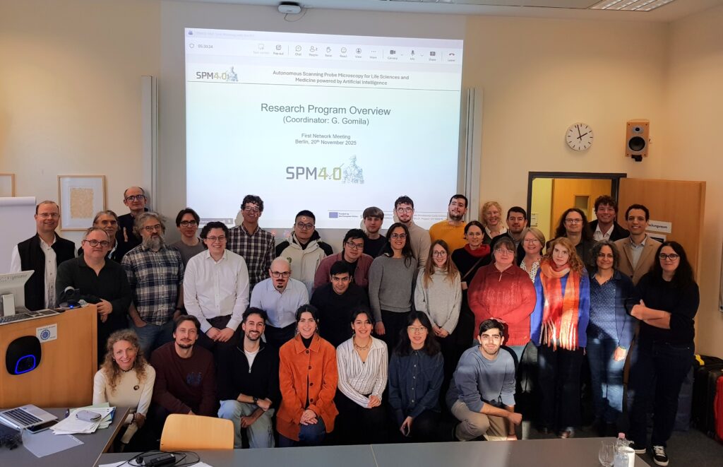
Berlín acull la reunió intermèdia i el primer curs de formació de la xarxa SPM4.0, coordinada per l’IBEC
El consorci SPM4.0 es va reunir a Charité-Berlín per celebrar el seu primer curs de formació i la seva reunió intermèdia per tal de reforçar la col·laboració científica i donar suport al creixement dels investigadors doctorals del projecte. Les sessions van oferir una visió general completa dels actuals avenços científics i de les futures activitats de formació. Aquesta fita va reforçar encara més la coordinació a tota la xarxa i va establir les bases per assolir els següents objectius del projecte.
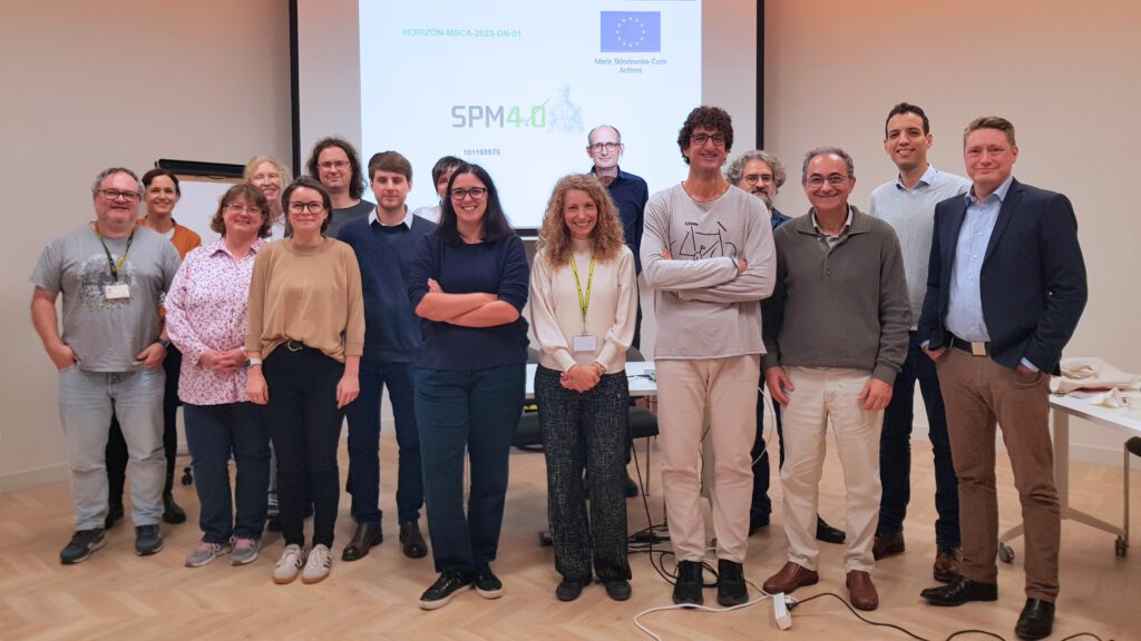
L’IBEC acull la reunió de llançament del projecte europeu SPM4.0
Investigadors de tot Europa es van reunir per a la reunió de llançament del projecte SPM4.0, una innovadora Xarxa Doctoral Marie Curie Skłodowska (MSCA-DN) dedicada a avançar en les capacitats de la microscòpia de sonda de rastreig (SPM) autònoma impulsada per intel·ligència artificial. L’esdeveniment, celebrat a l’IBEC, va marcar el començament d’una iniciativa amb visió de futur destinada a formar una nova generació d’investigadors i investigadores per ampliar els límits de la tecnologia dins dels camps de les ciències de la vida i la medicina.
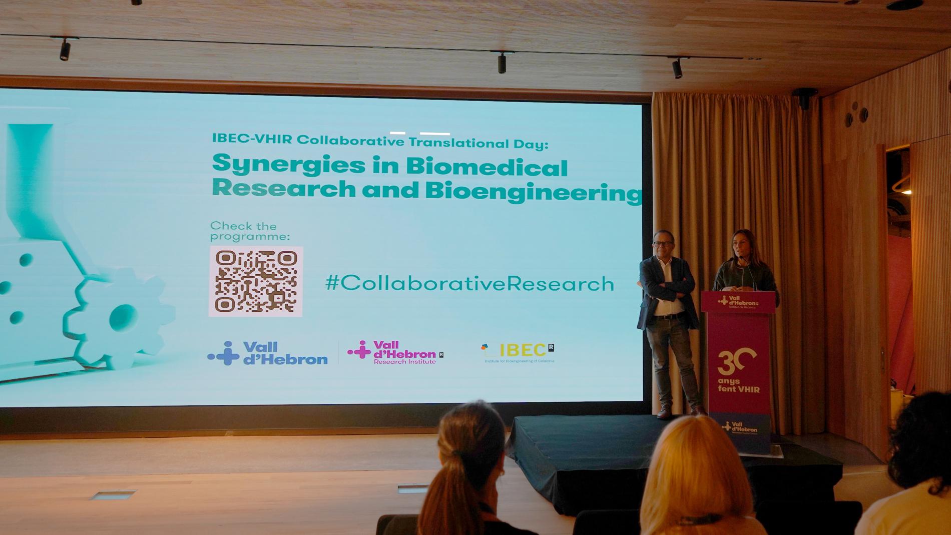
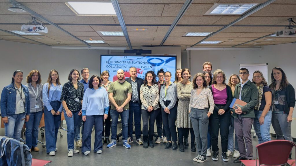
L’IBEC i el VHIR celebren una jornada de col·laboració per fomentar les sinergies
La 1a Jornada Col·laborativa Translacional entre el Vall d’Hebron Institut de Recerca (VHIR) i l’Institut de Bioenginyeria de Catalunya (IBEC), celebrada el 21 de novembre, ha estat una oportunitat per conèixer els projectes i les línies de recerca d’ambdues institucions i promoure la interacció entre els professionals.
L’IBEC i el Banc de Sang i Teixits enforteixen llaços amb una jornada de col·laboració translacional
L’IBEC i el Banc de Sang i Teixits han celebrat una jornada per explorar noves col·laboracions en bioenginyeria i medicina translacional. La trobada, celebrada ahir a l’IBEC, va destacar projectes innovadors, va presentar un programa de doctorat conjunt i va reforçar la connexió entre recerca biomèdica i aplicacions clíniques.
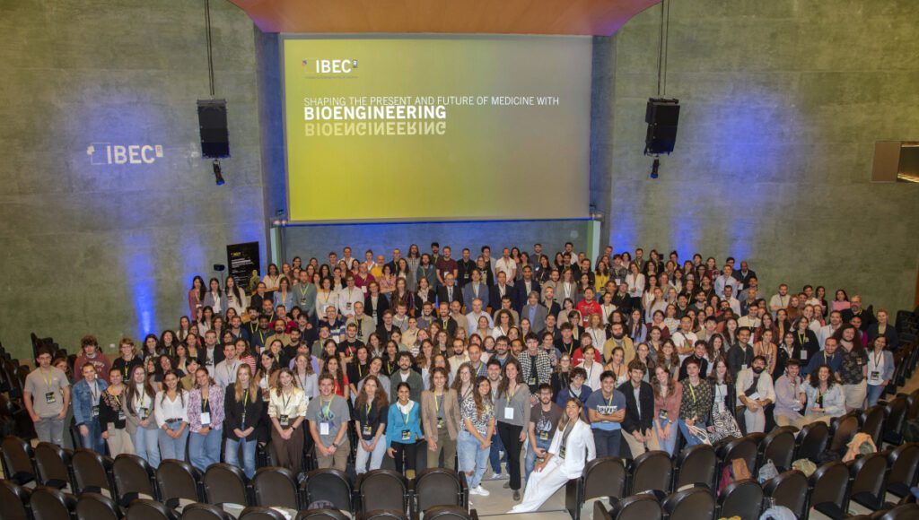
Bioenginyeria per a les teràpies emergents i avançades en el 17è Simposi de l’IBEC
El 17è Simposi anual de l’IBEC es va centrar en ‘Bioenginyeria per a les Teràpies Emergents i Avançades’, una de les àrees clau d’aplicació de l’IBEC. Van ser prop de 300 les persones assistents a l’esdeveniment, entre les quals es trobaven investigadors locals i internacionals. Un ambient multidisciplinari en el qual experts d’altres centres i la mateixa comunitat de l’IBEC van tenir l’oportunitat de presentar els seus projectes i intercanviar coneixement.
Dos projectes amb participació de l’IBEC seleccionats en la convocatòria de xarxes de doctorat MSCA
L’IBEC coordinarà SPM4.0 i participarà com a soci en ENTRY-DM, dos dels projectes seleccionats en la convocatòria 2023 de xarxes de doctorat dins del marc de les Accions Marie Skłodowska-Curie (MSCA). Gràcies a aquests dos projectes, l’IBEC incorporarà tres nous doctorands a la seva plantilla.
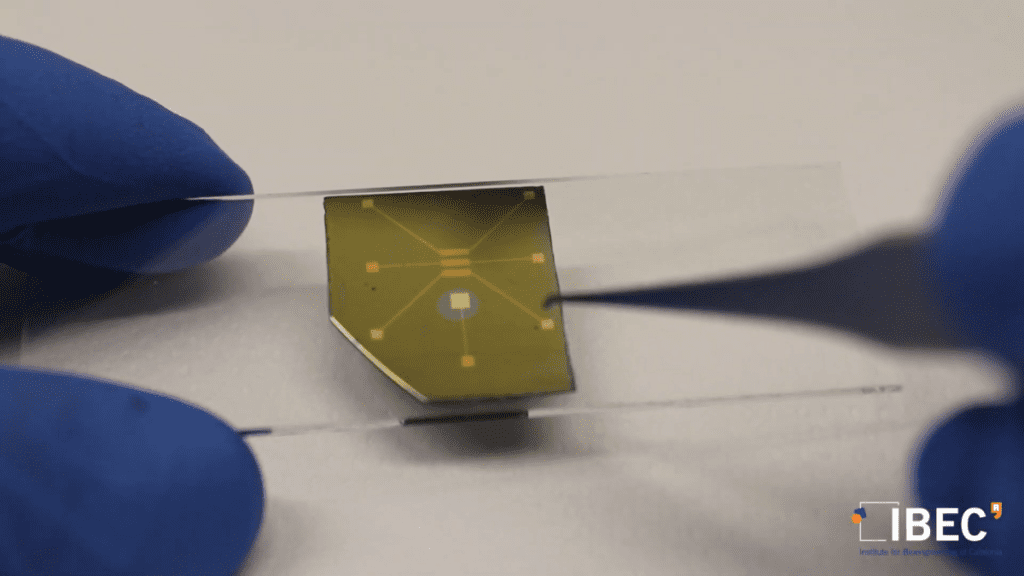
Nova metodologia per estudiar transistors orgànics en funcionament amb aplicacions en bioelectrònica
Un estudi liderat per l’IBEC ha aconseguit elaborar un mapa del potencial elèctric local al llarg de l’estructura de transistors orgànics usats en bioelectrònica que permet fer una avaluació detallada dels colls d’ampolla en el transport de càrrega. L’objectiu d’aquest estudi és aprofundir en la comprensió de les propietats del transport de càrrega en materials utilitzats en l’electrònica orgànica en contacte en medis líquids i millorar la seva aplicació en biosensors o enregistraments bioelèctrics.
Elisabeth Engel i Gabriel Gomila reben la distinció del programa ICREA Acadèmia
Els investigadors de l’IBEC Elisabeth Engel i Gabriel Gomila han estat guardonats amb la distinció “ICREA Acadèmia” que atorga la Institució Catalana de Recerca i Estudis Avançats (ICREA). Tant Engel … Read more

Research Assistant at the Nanoscale bioelectrical characterization group
Introduction to the vacant position: The Nanobioelec Group/Unit is looking for Research Assistant. The contract will be within the framework of the European Project PRINGLE, whose objective is to develop … Read more
JOBS

Senior Researcher position at the Nanoscale Bioelectrical Characterization research group
Ref: SR_GG // Deadline: 08/12/2024
Senior Laboratory Technician at the Nanoscale Bioelectric Characterization Research Group (Ref SRT_GG)
Ref: SRT_GG // Deadline: 20/06/2024
Post-doctoral researcher at the Nanobioelec Research Group (PD_GG)
Ref: PD_GG // Deadline: 15/01/2024

Predoctoral researcher at the Nanoscale Bioelectrical Characterization Research Group
Ref: FPI_GG /Deadline: 31/10/2023
Post-doctoral researcher at the Nanobioelec Research Group (PD_GG)
Ref: PD_GG // Deadline: 21/07/2023
Pre-doctoral researcher at the Nanobioelec Research Group (Phd_GG)
Ref: Phd_GG // Deadline 30/06/2023
Post-doctoral researcher at the Nanobioelec Research Group (Pd_GG)
Ref: Pd_GG // Deadline: 26/11/2023
Predoctoral researcher at the Nanoscale bioelectrical characterization group (PHD_GG)
Reference: PhD-GG / Deadline: 15/03/2023