
About
We aim at understanding how physical forces and molecular control modules cooperate to drive biological function.
We develop new technologies to map and perturb the main physical properties that determine how cells and tissues grow, move, invade and remodel.
By combining this physical information with systematic molecular perturbations and computational models we explore the principles that govern the interplay between chemical and physical cues in living tissues.
We study how these principles are regulated in physiology and development, and how they are derailed in cancer and aging.
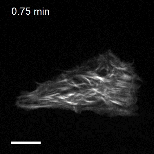
Making cellular forces visible
To study cell and tissue dynamics we develop new technologies to measure physical forces at the cell-cell and cell-matrix interface. By combining these technologies with computational analysis of cell shape and velocity we obtain a full experimental characterization of epithelial dynamics during tissue growth, wound healing and cancer cell invasion.
Tumour invasion by stromal forces
Cancer cell invasion and metastasis remain the leading cause of death in patients with cancer. Both processes are the result of a complex interaction between tumor cells and their microenvironment. One of our main lines of research is to study how tumours exploit the functions of non-cancer cells in their microenvironment to invade and metastasize. We focus on the interaction between epithelial cancer cells and Cancer Associated Fibroblasts (CAFs), the most abundant cell type in the tumour stroma.
Optogenetics to control cell mechanics
The recent development of optogenetic technologies offers promising possibilities to control signalling pathways with high spatiotemporal resolution. By expressing genetically encoded light-sensitive proteins, optogenetic technology enables the reversible perturbation of intracellular biochemistry with subcellular resolution. We have developed optogenetic tools based on controlling the activity of endogenous RhoA to upregulate or downregulate cell contractility and to control cell shape and mechanotransduction.
Collective durotaxis: a mechanism for cellular guidance by mechanical cues
Directed cell migration is one of the earliest observations in cell biology, dating back to the late XIX century. Also known as taxis, directed cell migration has been commonly associated with chemotaxis, i.e. the ability of a broad variety of cell types to migrate following gradients of chemical factors. We recently demonstrated a new mode of collective cell guidance by mechanical cues, called collective durotaxis. This new migration mode emerges only in cell collectives and, strikingly, does not require isolated cells to exhibit gradient sensing.
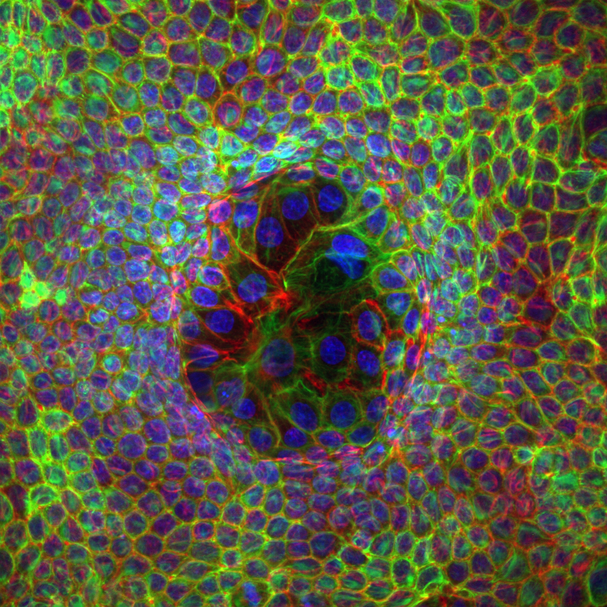
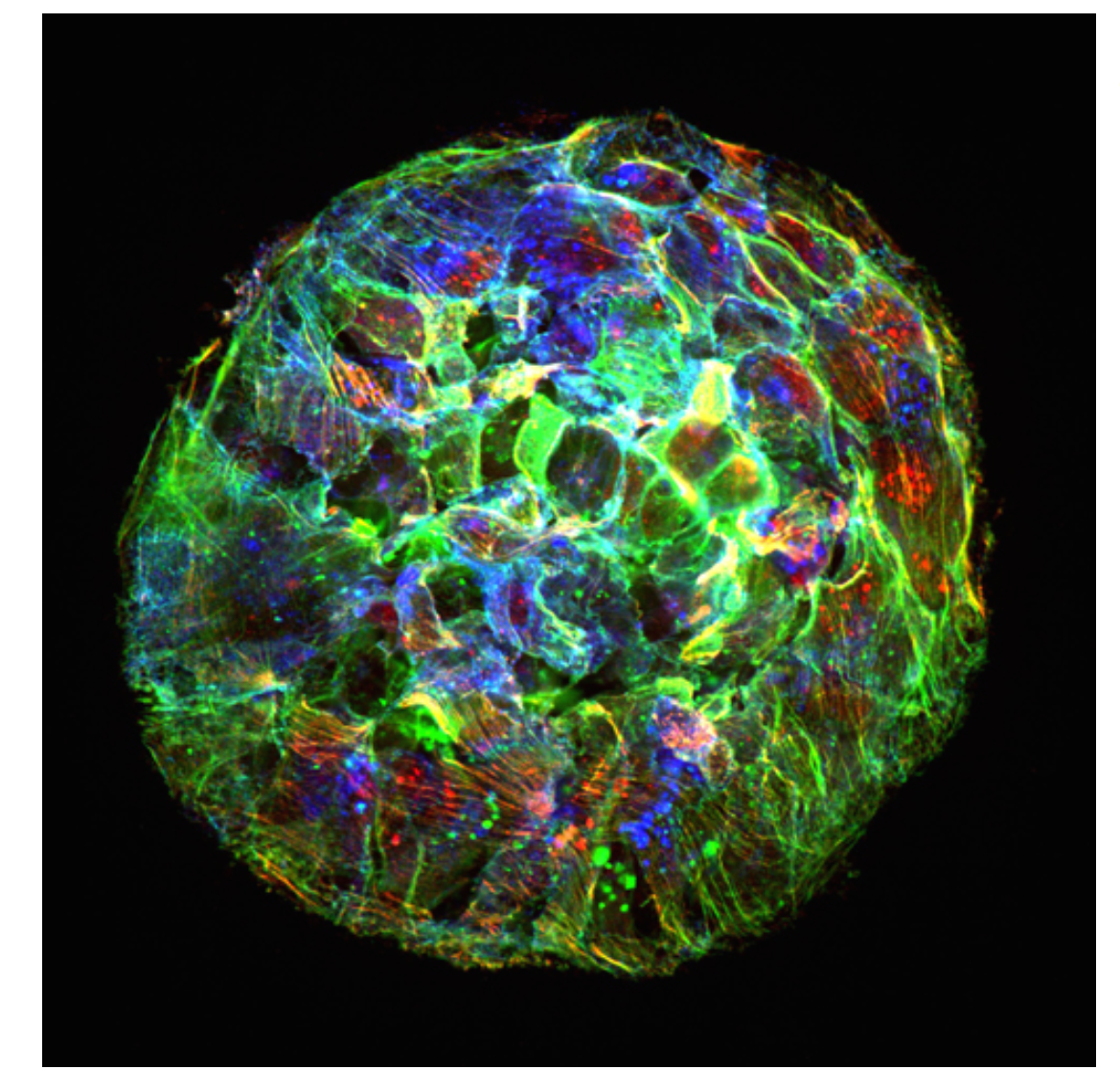
Organoid mechanobiology
Organoids are large multicellular structures that self-organize in vitro and maintain a similar organization and functionality than the organ from which they are derived. Organoids from many organs have now been obtained from embryonic stem cells, induced pluripotent stem cells and organ progenitors. We use intestinal and kidney organoids to study how epithelia adopt three-dimensional shapes that closely resemble their structure in vivo. We also use organoids grown from primary tumors to understand how epithelial structure and function are lost with disease progression.
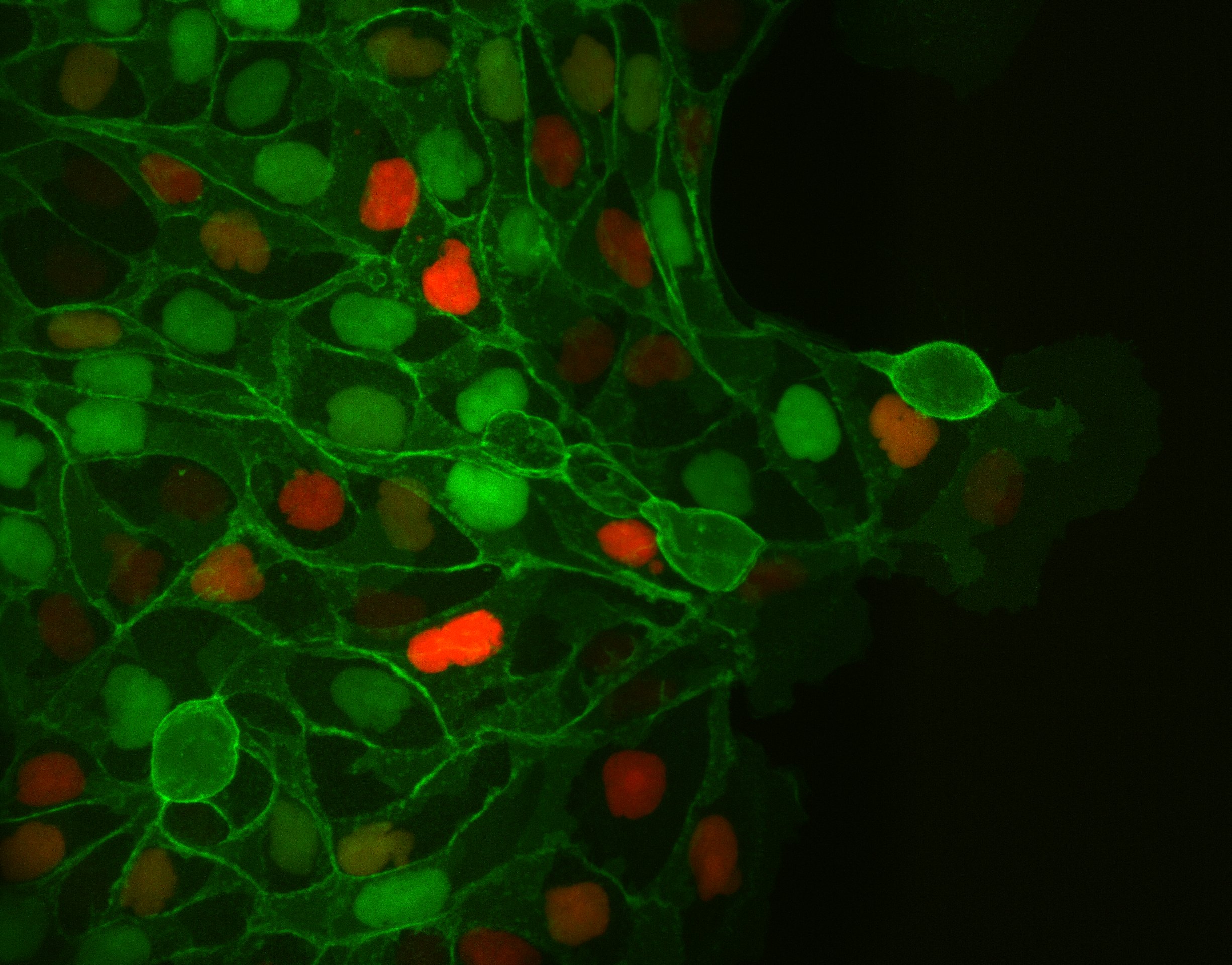
Engineering epithelial shape and mechanics from the bottom up
We develop new approaches to engineer epithelia in 3D. Using these approaches, we study the principles that govern the emergence of tissue shape from the bottom up. We recently found that epithelial sheets can stretch up to four times their initial area without breaking, and that they are able to recover their initial size in a fully reversible way when unstretched. Surprisingly, some cells in the tissue barely stretch, while others become ‘superstretched’, increasing their area more than ten times. We call this phenomenon ‘active superelasticity’.
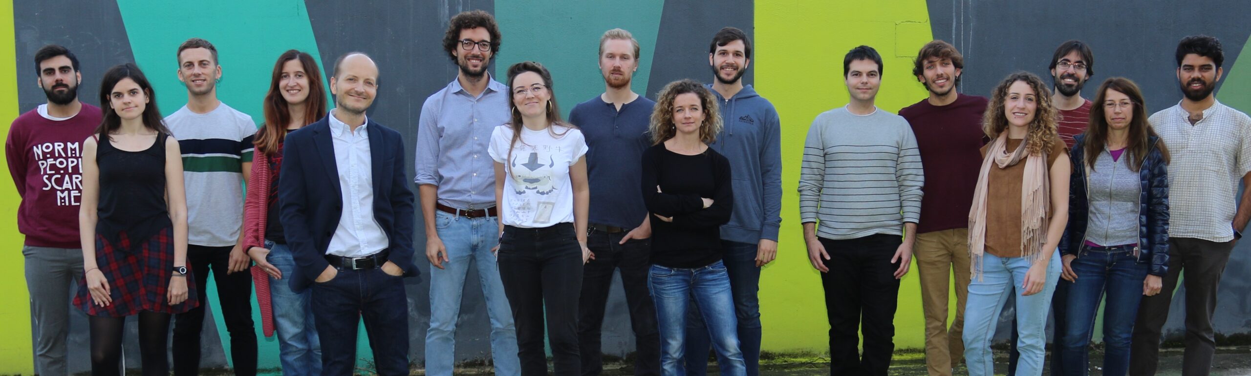
Staff
[br]
 ibecbarcelona.eu
Projects
| NATIONAL PROJECTS | FINANCER | PI |
|---|---|---|
| mGRADIENTMecanobiología de la migración colectiva durante la haptotaxis y la durotaxis: aplicación a los organoides intestinales (2019-2022) | MICIU Generación Conocimiento: Proyectos I+D | Xavier Trepat |
| DYNAGELHidrogeles biocompatibles con rigidez dinámicamente ajustable para estudiar la mecanobiología de células y tejidos (2019-2022) | MICIU Retos investigación: Proyectos I+D | Raimon Sunyer |
| INTERNATIONAL PROJECTS | FINANCER | PI |
|---|---|---|
| EpiFold Engineering epithelial shape and mechanics: from synthetic morphogenesis to biohybrid devices (2021-2025) | European Commission, ERC-AdG | Xavier Trepat |
| The role of intermediate filaments in stress resistance in 3D epithelial structures (2021-2023) | Deutsche Forschungsgemeinschaft (DFG), Walter Benjamin-Programme | Tom Golde |
| Mechano·Control Mechanical control of biological function (2017-2022) | European Commission, FET Proactive | Xavier Trepat |
| Control of cell collective flows and tissue folding by means of surface patterns (2021-2022) | Human Frontier Science Program, HFSP Beca postdoctoral | Pau Guillamat |
| PRIVATELY-FUNDED PROJECTS | FINANCER | PI |
|---|---|---|
| Mech4Cancer · Enabling technologies to map nuclear mechanosensing: from organoids to tumors (2020-2023) | Obra Social La Caixa: Health Research Call | Xavier Trepat |
| T cell exclusion during cancer immune evasion and immunotherapy failure: cell types, transcriptional programs and biomechanics (2020-2023) | Fundació La Marató de TV3 | Xavier Trepat |
| Joint Programme Healthy Ageing | Obra Social La Caixa | Xavier Trepat |
| Understanding and measuring mechanical tumor properties to improve cancer diagnosis, treatment, and survival: Application to liquid biopsies (2017-2022) | Obra Social La Caixa | Xavier Trepat |
| FINISHED PROJECTS | FINANCER | PI |
|---|---|---|
| OPTOLEADER Optogenetic control of leader cell mechanobiology during collective cell migration (2019-2021) | European Commission, MARIE CURIE – IF | Leone Rossetti |
| MECHANOIDS Probing and controlling the three-dimensional organoid mechanobiology (2019-2021) | European Commission, MARIE CURIE – IF | Manuel Gómez |
| TensionControl Multiscale regulation of epithelial tension (2015-2020) | European Commission, ERC – CoG | Xavier Trepat |
| El mecanoma de la adhesión epitelial: mecanismos de detección, resistencia y transmisión de fuerzas intercelulares | MINECO, I+D-Investigación fundamental no orientada | Xavier Trepat |
| MICROGRADIENTPAGE Micro Gradient Polyacrylamide Gels for High Throughput Electrophoresis Analysis | European Commission, ERC-PoC | Xavier Trepat |
| GENESFORCEMOTION Physical Forces Driving Collective Cell Migration: from Genes to Mechanism | European Commission, ERC-StG | Xavier Trepat |
| CAMVAS Coordination and migration of cells during 3D Vasculogenesis (2014-2017) | European Commission, MARIE CURIE – IOF | Xavier Trepat |
| DUROTAXIS Mecanobiología de la durotaxis: de las células aisladas a los tejidos | MINECO, Proyectos I+D Excelencia | Xavier Trepat |
Publications
Check for more detailed information on the outputs of the Group at IBEC CRIS portal.
Publications list:
Equipment
- Soft Lithography
- Micro/Nano fabrication
- Cell stretching
- Live Confocal Microcopy
- Magnetic Tweezers
- Magnetic Twisting Cytometry
- Monolayer stress microscopy
- Traction microscopy
Collaborations
- Julien Colombelli / Eduard Batlle
Institute for Research in Biomedicine (IRB) Barcelona - Marino Arroyo
Universitat Politècnica de Catalunya, Barcelona - Guillaume Charras / Roberto Mayor
University College London, UK - Erik Sahai
Cancer Research, UK - Benoit Ladoux
Université Paris 7, France - Jim Butler & Jeff Fredberg
Harvard University, Boston - Danijela Vignjevic
Institut Curie, Paris - Jonel Trebicka
Department of Internal Medicine I, University Hospital Frankfurt - Eduard Batlle
Institute for Research in Biomedicine (IRB) Barcelona
News
Se celebra a Barcelona la quarta edició de la Conferència EMBL-IBEC
La quarta edició de la Conferència EMBL-IBEC organitzada per l’Institut de Bioenginyeria de Catalunya (IBEC) i el Laboratori Europeu de Biologia Molecular (EMBL) va girar al voltant del modelatge de malaltia del desenvolupament i medicina regenerativa, i va reunir aquesta setmana al PRBB de Barcelona prop de 130 experts i expertes internacionals en el camp de la bioenginyeria.
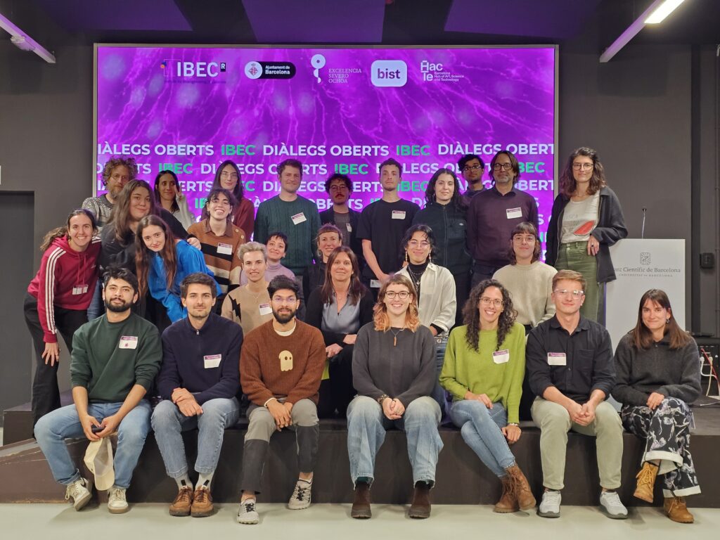
L’IBEC celebra amb èxit la Hackathon del seu nou programa de cocreació “Diàlegs Oberts”
L’Institut de Bioenginyeria de Catalunya va reunir ahir una vintena d’artistes i científics i científiques en una Hackathon de cocreació dins el marc de la seva nova iniciativa “Diàlegs Oberts”. Com a resultat de la jornada es van seleccionar dues parelles artista-científic que, durant els pròxims cinc mesos, col·laboraran per desenvolupar sengles projectes basats en la recerca de l’IBEC.
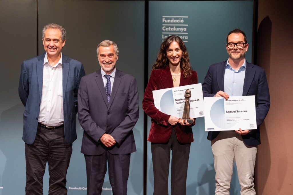
Una investigació de l’IBEC guanya el 3r Premi Vanguardia de la Ciència 2025
Un estudi liderat des de l’IBEC ha estat guardonat amb el 3r Premi Vanguardia de la Ciència 2025. La investigació, que té com a primera autora Meritxell Serra-Casablanca i com a líder Samuel Sánchez Ordoñez, proposa una teràpia innovadora contra el càncer de bufeta basada en nanorobots carregats amb radiofàrmacs, capaços de desplaçar-se per la bufeta aprofitant l’orina com a font d’energia.
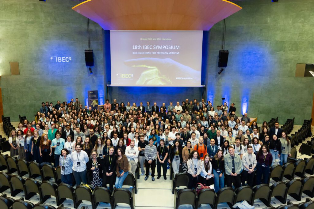
Bioenginyeria per a la medicina de precisió en el 18è Simposi de l’IBEC
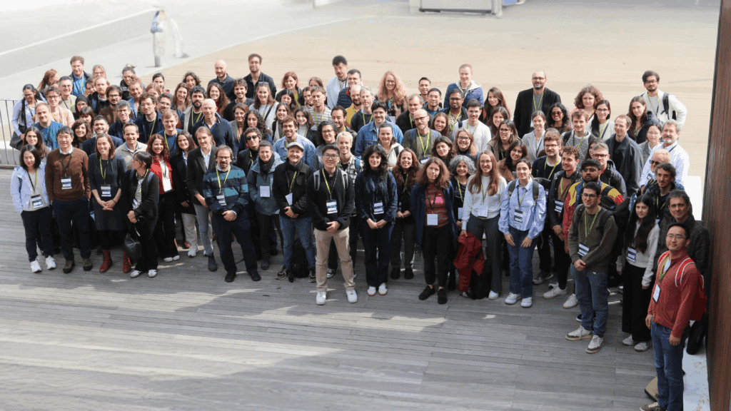
El 18è Simposi anual de l’IBEC es va centrar en ‘Bioenginyeria per a la Medicina de Precisió’, una de les àrees clau d’aplicació de l’IBEC. Van ser prop de 300 les persones assistents a l’esdeveniment, entre les quals es trobava personal investigador local i internacional. Un ambient multidisciplinari en el qual experts i expertes d’altres centres i la mateixa comunitat de l’IBEC van tenir l’oportunitat de presentar els seus projectes i intercanviar coneixement.
L’IBEC i l’EMBL Barcelona coorganitzen una jornada de col·laboració per explorar sinergies
L’Institut de Bioenginyeria de Catalunya (IBEC) i el Laboratori Europeu de Biologia Molecular (EMBL) han celebrat avui una jornada de “matchmaking”. L’esdeveniment ha reunit investigadors i investigadores destacades d’ambdós centres per tal de fomentar la creació de noves connexions i promoure el diàleg científic.
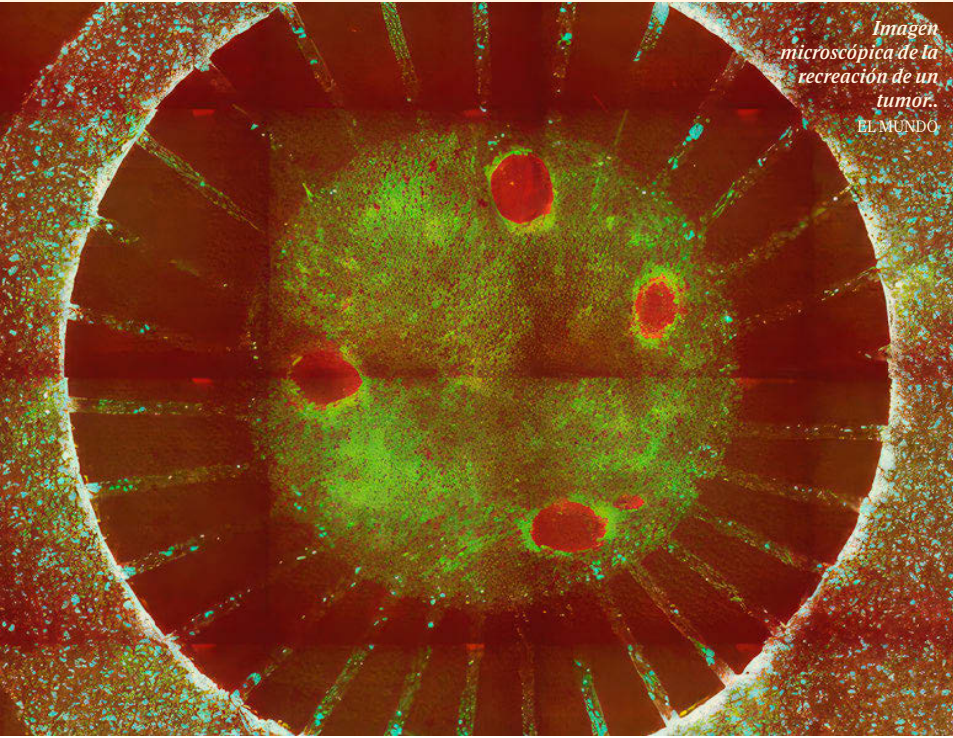
El Mundo: Un ‘gemelo’ de laboratorio para estudiar la eficacia de los tratamientos contra el cáncer
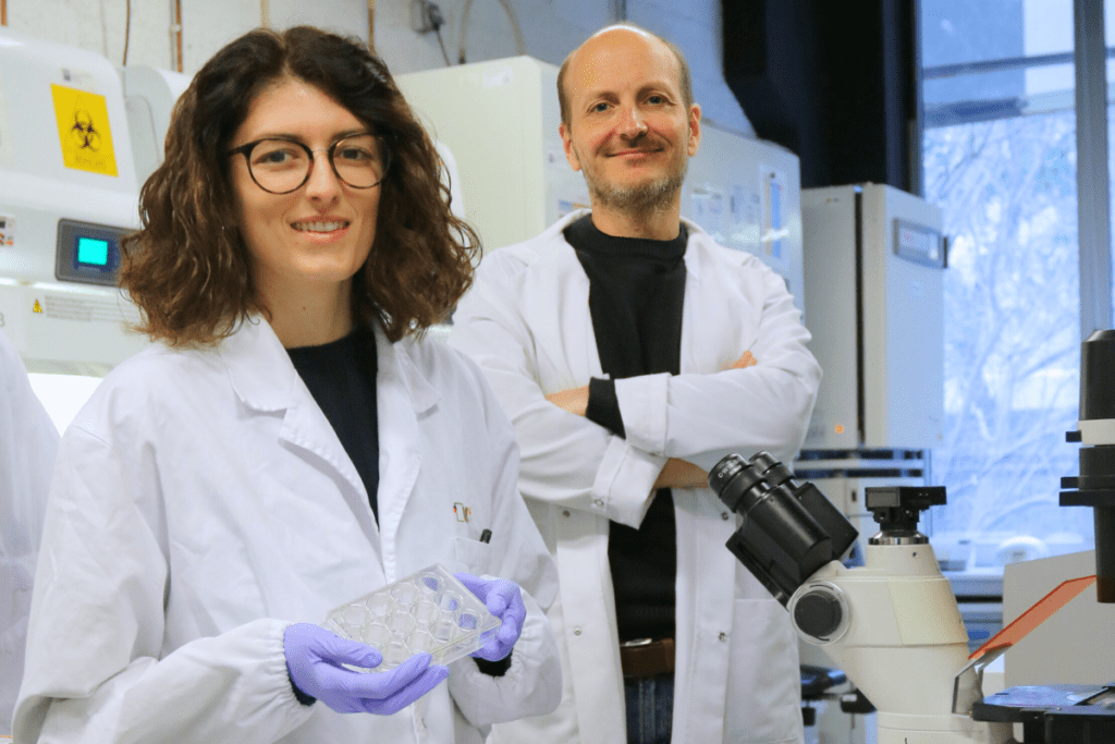
Científicos del IBEC, liderados por Xavier Trepat, han desarrollado MIRO, un dispositivo que recrea el entorno tumoral para estudiar su respuesta a tratamientos. Publicado en Nature Communications, permite analizar por … Read more
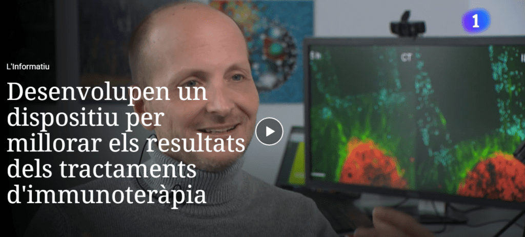
RTVE: Desarrollan un dispositivo para mejorar los resultados de tratamientos de inmunoterapia
Investigadores de l’Hospital del Mar y del IBEC están desarrollando un dispositivo para mejorar los resultados de los tratamientos de inmunoterapia contra el cáncer. Este dispositivo podría optimizar la eficacia … Read more
La Vanguardia: Investigadores catalanes recrean de forma fidedigna el ambiente tumoral para atacar el cáncer
Investigadores del IBEC y del Hospital del Mar han desarrollado MIRO, un dispositivo que recrea fielmente el entorno tumoral con células de pacientes. Esta innovadora tecnología permite estudiar la interacción … Read more
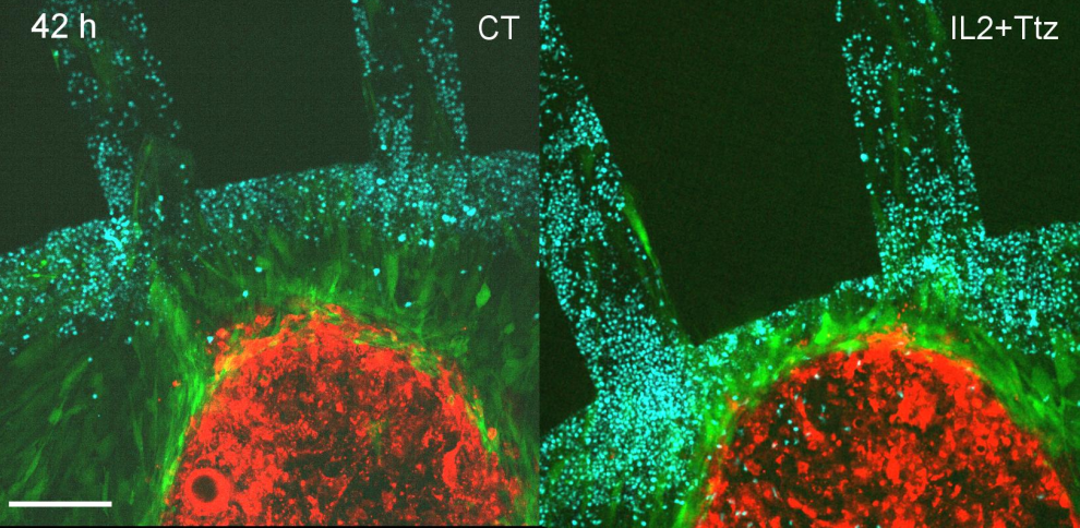
Desenvolupen un dispositiu que replica els tumors per estudiar l’eficàcia de tractaments amb immunoteràpia
El Micro Immune Response On chip (MIRO) permet replicar els tumors i el seu entorn, per conèixer la seva resposta als tractaments basats en inmunoteràpia. El dispositiu, que ja s’ha provat amb èxit en mostres de càncer de mama, pot ser clau per desenvolupar nous tractaments i determinar quina és la teràpia més adequada per a cada pacient de manera personalitzada. El treball, publica Nature Communications, és fruit de la col·laboració entre l’Institut de Bioenginyeria de Catalunya i l’Institut de Recerca de l’Hospital del Mar.
Jobs
Postoctoral Researcher at the Integrative Cell and Tissue Dynamics Research Group
Ref: PR-XT // Deadline: 21/12/2025
Laboratory Technician at the Integrative Cell and Tissue Dynamics Research Group
Ref: LA-XT // Deadline: 24/11/2025
Research assistant at the Integrative Cell and Tissue Dynamics Research Group
Ref: RA-XT // Deadline: 17/10/2025
Research Training at the Integrative Cell and Tissue Dynamics Research Group
Ref: RT_XT // Deadline: 15/08/2025
Research assistant at the Integrative Cell and Tissue Dynamics Research Group
Ref: RA-XT // Deadline: 24/07/2025
Research Assistant at the Integrative Cell and Tissue Dynamics Research Group
Ref: RA_XT//Deadline: 5/11/2024
Postdoc at the Integrative Cell and Tissue Dynamics Research Group
Ref: XT-PD/Deadline: 18/12/2023
Research assistant at the Integrative Cell and Tissue Dynamics Research Group
Ref: RA_XT/Deadline: 15/06/2023