

A new technique developed at the Institute for Bioengineering for Catalonia (IBEC) makes it possible to classify the quality of embryos faster and twice as accurately as expert embryologists. The technology, called “METAPHOR”, uses imaging and artificial intelligence to analyse the metabolism of embryos and oocytes. This technique promises to drastically reduce the time and treatment cycles needed to achieve pregnancy through in vitro fertilisation, minimising the emotional and financial burden. on patients.
A new technology developed by the “Bioengineering in Reproductive Health” team at the Institute for Bioengineering of Catalonia (IBEC) is able to visualise the metabolism of embryos obtained through in vitro fertilisation in order to decide which are most likely to implant correctly in the uterus and reaching full-term. It is a more accurate and reliable technique than traditional methods.
This new technology will help to increase the probability of success in assisted reproduction processes, reducing the so-called ‘time to pregnancy’ and the economic and psychological burden on patients.
Samuel Ojosnegros
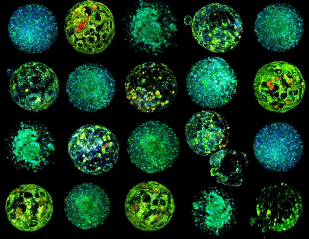
The revolutionary method, called METAPHOR”, generates 3D images that reveal the colours present in the embryo in a completely non-invasive way. Certain naturally fluorescent compounds in the embryo’s metabolism are also key to processes such as cellular respiration or nutrient consumption, making METAPHOR a reliable way to monitor the embryo’s health.
“This new technology will help to increase the probability of success in assisted reproduction processes, reducing the so-called ‘time to pregnancy’ and the economic and psychological burden on patients,” says Samuel Ojosnegros, principal investigator at IBEC and leader of the study.
We are able to assess the loss of oocyte quality associated with the loss of fertility with age. This would be a breakthrough in the management of fertility donation and preservation.
Anna Seriola
The paper, published in the prestigious journal PNAS, describes how, in studies with mice, they were able to double the success rate in selecting viable embryos compared to embryologists using traditional microscopy. In addition to embryo analysis, the method is highly accurate in analysing oocyte metabolism, allowing the most suitable oocytes to be selected for in vitro fertilisation. To do this, they compared oocytes from young and older females, as age is known to be crucial for oocyte viability. The METAPHOR system discriminated between young and non-young oocytes with 96% accuracy and was able to predict which would develop into viable embryos with over 80% accuracy, an unprecedented figure in the field.
“We are able to assess the loss of oocyte quality associated with the loss of fertility with age. We look for so-called ‘molecular signatures’, characteristics of the cells that are associated with this loss of fertility, such as the distribution of mitochondria. From this information, we can predict which oocytes will develop and which will not. This would be a breakthrough in the management of fertility donation and preservation,” says Anna Seriola, senior researcher in the Ojosnegros group and author of the study.
We trained an artificial intelligence tool capable of analysing and classifying hundreds of images in a few minutes.
Albert Parra
The technological basis of METAPHOR uses artificial intelligence methods to analyse metabolic images obtained by hyperspectral microscopy. “Using hyperspectral microscopy, we acquire hundreds of images containing complex information of many mixed metabolites from embryos and oocytes. To analyse them, we trained an artificial intelligence tool capable of analysing and classifying these images in a few minutes,” says Albert Parra, a researcher in the Ojosnegros group and first author of the study.
The power and safety of this new method position it as a revolutionary tool for assessing oocytes and embryos based on their physiology. Researchers are currently fine-tuning the technology to evaluate human embryos and have established a spin-off company to bring the technology to assisted reproduction clinics in the coming years.
Referenced Article:
Albert Parra, Denitza Denkova, Xavier P. Burgos-Artizzu, Ester Aroca, Marc Casals, Irene Oliver-Vila, Miguel Ares, Anna Ferrer-Vaquer, Enric Mestres, Mònica Acacio, Nuno Costa-Borges, Elena Rebollo, Hsiao Ju Chiang, Scott E. Fraser, Francesco Cutrale, Anna Seriola, and Samuel Ojosnegros. METAPHOR: Metabolic Evaluation through Phasor-based Hyperspectral Imaging and Object Recognition for Mammalian Blastocysts and Oocytes. PNAS (2024). DOI: https://doi.org/10.1073/pnas.2315043121


