

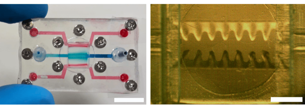
The innovative device contains a 3D bioprinted channel with structures that mimic intestinal villi and reproduce the compartments of the intestinal mucosa. For the first time, electrodes have been incorporated into the system to monitor the formation of the intestinal barrier in real time. The device is highly versatile and has potential applications in disease modelling and drug screening.
The most innovative thing is that we combine bioprinting directly on the fluidic chip.
Elena Martínez Fraiz
The Biomimetic Systems for Cell Engineering group at the Institute for Bioengineering of Catalonia (IBEC), in collaboration with the Biomedical Applications Group (GAB) of the Institute of Microelectronics of Barcelona (IMB-CNM), has developed an advanced model of gut-on-chip. It is an innovative device that contains a 3D bioprinted channel with structures that mimic the intestinal villi and reproduces the epithelial and stromal compartments of the intestinal mucosa. Electrodes have been incorporated into this model to monitor the formation of the intestinal barrier in real time. The device is highly versatile and has potential applications in disease modelling and drug screening.
“In our study, we integrate the soft part, made of hydrogels with cells, with a suitable morphology. The most innovative thing is that we do this by combining bioprinting directly on the fluidic chip,” explains Elena Martínez Fraiz, IBEC Senior Researcher, Associate Professor at the University of Barcelona (UB) and leader of the study.
In the near future we could develop a model to study inflammatory bowel disease or disorders related to bacterial overpopulation.
Núria Torras Andrés
The team is already working on making the system even more complex: “Our goal is to be able to simulate conditions that are closer to reality and, in the near future, to develop a model to study, for example, inflammatory bowel disease or disorders related to bacterial overpopulation,” says Núria Torras Andrés, senior researcher in Martínez’s group and author of the study.
The long-term goal is to obtain models that are more realistic and closer to human physiology, in order to replace animal experiments and make medicine much more personalised.
María García Díaz
In the field of organ-on-chip, conventional gut-on-chip models have struggled to reproduce the complexity of the human gut, particularly in fully representing its essential components. The gut is composed of several layers, including the epithelium, which forms the inner surface, and the stroma, a connective tissue that supports the epithelium and maintains the integrity of the intestinal barrier. The lack of adequate representation of these components has limited the development of effective models. However, new technologies such as 3D bioprinting and the use of hydrogels as extracellular matrix analogs are opening up new possibilities in the field of organ-on-chip.
“The long-term goal is to obtain models that are more realistic and closer to human physiology, in order to replace animal experiments and make medicine much more personalised,” explains María García Díaz, senior researcher in Martínez’s group and author of the study.
Referenced article:
Daniel Vera, María García-Díaz, Núria Torras, Óscar Castillo, Xavi Illa, Rosa Villa, Mar Alvarez and Elena Martinez. A 3D bioprinted hydrogel gut-on-chip with integrated electrodes for transepithelial electrical resistance (TEER) measurements. Biofabrication (2024). DOI: https://doi.org/10.1088/1758-5090/ad3aa4


