An IBEC-led study describes the development of an innovative method to control the formation of crypt-like structures and villi in the intestine using a contact protein printing technique. This model will make it possible to study in detail key processes such as cell regeneration or changes associated with diseases such as cancer and chronic inflammatory disorders.

The method developed by IBEC’s Biomimetic Systems for Cell Engineering group is based on the imprinting of defined patterns of key proteins, such as Wnt3a and EphrinB1, onto a basement membrane. These proteins are essential for the organisation and differentiation of intestinal epithelial tissue. Using this technique, the researchers were able to control how and where structures such as crypts and villi form in the intestine. The system also allows them to study the role of each of these proteins individually and in a controlled manner.
What we achieve with our method, which is based on contact printing of proteins, is to control how and where these structures are formed.
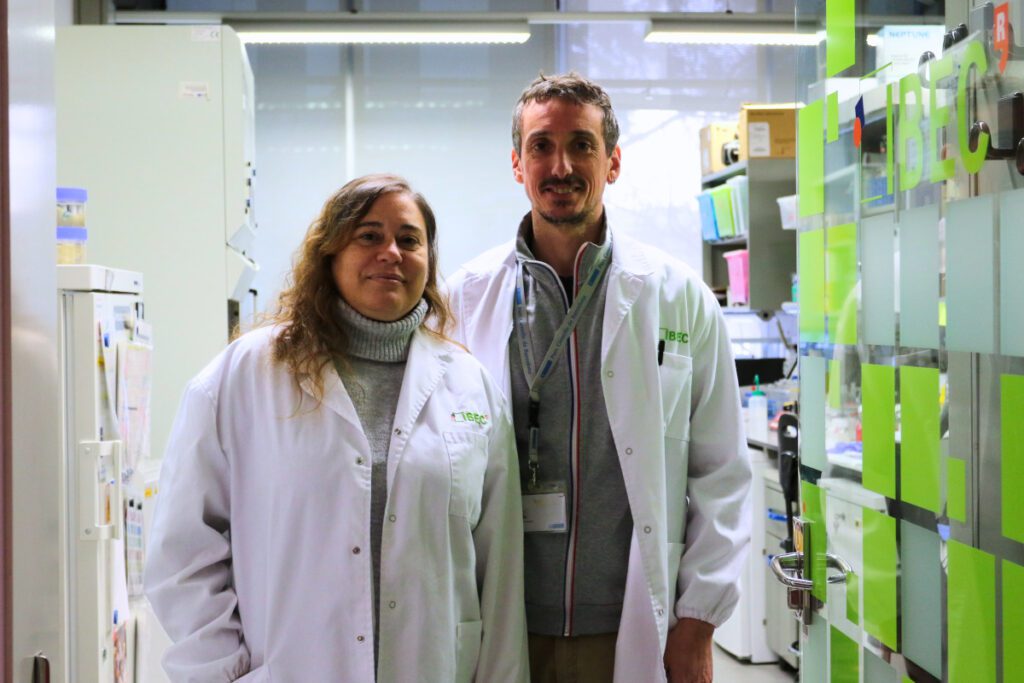
Jordi Comelles
‘The cells we are working with self-organise into distinct compartments that precisely replicate intestinal structures. What we achieve with our method, which is based on contact printing of proteins, is to control how and where these structures are formed. We do this by arranging these proteins in specific patterns, such as circles or holes,’ explains IBEC senior researcher Jordi Comelles, associate professor at the University of Barcelona (UB) and co-author of the study.
This innovative method also allows the individual analysis of the factors involved in the organisation and functioning of the intestine, revealing their role in key processes such as cell proliferation and differentiation. “For example, we have observed that exogenous Wnt3a can reduce the production of the same factor at the endogenous level, which opens up new possibilities for manipulating these signalling pathways,” adds Comelles.
This model will allow us to study in detail key processes such as cell regeneration or changes associated with diseases such as cancer and chronic inflammatory diseases.
Elena Martínez
This approach allows to control how intestinal cells cluster, depending on the size and arrangement of the Wnt3a patterns. ‘Our aim was to create a system that more closely mimics human intestinal tissue. This model will allow us to study in detail key processes such as cell regeneration or changes associated with diseases such as cancer and chronic inflammatory diseases,’ says Elena Martínez Fraiz, IBEC senior researcher, UB associate professor and leader of the study.
The team also used computer models to simulate the interactions between signalling pathways, providing a more detailed view of the processes involved in cellular organisation. This breakthrough not only improves our understanding of gut biology, but also opens up new opportunities to test drugs, study diseases in a controlled environment and develop more effective treatments.
This work is part of Enara Larrañaga’s doctoral thesis in Martínez’s group at IBEC. The research has also involved the collaboration of IBEC’s Bioengineering in Reproductive Health group, led by Samuel Ojosnegros; the Centro de Investigación Biomédica en Red – Bioingeniería, Biomateriales y nanomedicina (CIBER-BBN); the European Molecular Biology Laboratory (EMBL) in Barcelona; and the Institute for Research in Biomedicine (IRB Barcelona).
Referenced article:
Enara Larrañaga, Miquel Marin-Riera, Aina Abad-Lázaro, David Bartolomé-Català, Aitor Otero, Vanesa Fernández-Majada, Eduard Batlle, James Sharpe, Samuel Ojosnegros, Jordi Comelles & Elena Martínez. Long-range organization of intestinal 2D-crypts using exogenous Wnt3a micropatterning. Nature Communications (2025). DOI: https://doi.org/10.1038/s41467-024-55651-7


