


This new approach has been tested in patients and combines thoracic bioimpedance and electrical and mechanical respiratory muscular signals allowing to measure the level of pulmonary function in a non-invasive and quantitative way, reducing the inconveniences for the patients.
Chronic Obstructive Pulmonary Disease (COPD) is a common disease occurring in adults characterized by breathing problems and poor airflow. It is the fifth leading cause of death worldwide and a major cause of chronic morbidity and mortality. Currently, to assess the pulmonary function, patients are subjected to tests that rely in maximal manoeuvres and to an increase in the overall inspiratory effort needed to breath in order to quantify the level of airflow limitation. Moreover, to fully evaluate the patient, the result of the tests must be completed with information from questionnaires, what includes a certain grade of subjectivity and non-quantifiable data that difficult an objective assessment.
Now, a team of researchers led by Raimon Jané, Leader of the Biomedical Signal Processing and Interpretation Group (BIOSPIN) at IBEC and UPC Professor, in collaboration with imec researchers from The Netherlands and Belgium (prof. Francky Catthoor), and the Ziekenhuis Oost-Limburg Hospital in Belgium, publish in the IEEE Transactions on Biomedical Engineering Journal, a new approach to evaluate COPD. The novel methodology, non–invasive and quantifiable, was applied to COPD patients and is based on the combination of bioimpedance and myographic signals to measure the contribution of inspiratory muscle activity into pulmonary ventilation. This proposal is one of the relevant outcomes of the joint project “Evaluation of non-invasive indices for respiratory monitoring, focusing on the applicability at home” by the IBEC and imec, and the project “Smart health ecosystem (tools, apps and devices) for personalized medicine and Healthcare in Respiratory diseases and Sleep disorders” (SappHiRES), led by prof. Jané.
A more comfortable and non-invasive method to monitor pulmonary diseases
Respiratory muscle disfunction occurs very often in COPD patients and contribute to their breathlessness feeling. This dysfunction is measured by the inspiratory muscle activity and global respiratory output, and the data are compared to healthy people to evaluate the effectiveness of respiratory muscles in ventilation. An example of a current procedure to evaluate respiratory muscle disfunction is an invasive measurement of the neural respiratory effort of the crural part of the diaphragm using a catheter, , which is uncomfortable for the patient, requires an elaborated clinical assistance and represents a disadvantage for systematic disease monitoring.
Some non–invasive procedures to assess pulmonary dysfunction include the measurement of physiological signals by surface sensors attached to the patient’s skin, such as bioimpedance and myographic signals. On one hand, bioimpedance measures the ability of a biological tissue to impede an electric current. On the other hand, electromyographic (EMG) and mechanomyographic (MMG) signals measure the electrical and mechanical activity of the muscles respectively, in the case of this study, the lower chest wall. These three non-invasive measures have been demonstrated to be capable to assess respiration independently but have never been combined to give a broader picture of pulmonary dysfunctions.
The novelty of the approach presented in this work relies on the simultaneous and non-invasive measurements of bioimpedance and electrical and mechanical muscular signals from the lower intercostal spaces, to investigate ventilatory responses during COPD patients’ breathing.
Dolores Blanco-Almazán, first author of the work
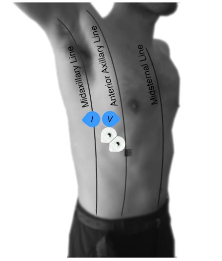
In this study, bioimpedance, EMG and MMG signals were acquired using a wearable research device and a wired standard acquisition system. Both bioimpedance and EMG signals were captured by surface electrodes whereas MMG signals were recorded by an accelerometer, all adhesively fixed on the thoracic region of the patients who stayed comfortably seated in an upright position during all the procedure. The new version of the acquisition system used, a system on chip (SoC), allows bioimpedance, EMG and MMG simultaneous recording applying a multimodal approach to reduce the intrusiveness, what reinforces the patient-friendly breathing monitoring and its potential ambulatory or home use.
Image: representation of the location of the electrodes (blue and white patches) and accelerometer (small black square).
New indexes to quantify respiratory muscle dysfunction
Researchers propose two new indexes to quantify respiratory muscle dysfunction in COPD patients: the BEr and BMr ratios. They evaluate the variation of bioimpedance and the electrical and mechanical activity during each respiratory cycle and quantify the contribution of lower chest wall inspiratory muscle activation to ventilation, that is, the ratio of bioimpedance to EMG (BEr) and to MMG (BMr).
These new indexes consider different parameters related to breathing pattern and showed significant differences between mild and severe COPD patients, with mild patients presenting a higher contribution of the inspiratory muscles to global respiratory ventilation than severe ones, that is, severe COPD patients need significantly higher electrical and mechanical muscle activity levels to breath. Thus, both bioimpedance and myographic signals are potential alternatives to the traditional invasive and obtrusive methods in characterizing respiratory diseases.
This work evaluates the contribution of the muscle activity into global respiratory ventilation by the use of two new indexes and validate the combined use of bioimpedance and myographic signals to provide useful parameters to noninvasively assess COPD.
Raimon Jané
The new procedure proposed in this study opens the door to reinforce the combination of bioimpedance and myographic signals to monitor respiratory diseases in a noninvasive and fully quantifiable way, helping to evaluate improvement or deterioration of pulmonary dysfunction in COPD patients.
Reference article: Dolores Blanco-Almazán, Willemijn Groenendaal, Manuel Lozano-García, Luis Estrada-Petrocelli, Lien Lijnen, Christophe Smeets, David Ruttens, Francky Catthoor and Raimon Jané. Combining Bioimpedance and Myographic Signals for the Assessment of COPD during Loaded Breathing. IEEE Transactions on Biomedical Engineering (2021), 68 (1), 298-307.
Blanco-Almazán, M. Lozano-García, L. Estrada-Petrocelli and R. Jané are members of the BIOSPIN (IBEC) and the Biomedical Research Networking Center in Bioengineering, Biomaterials and Nanomedicine (CIBER- BBN).


