

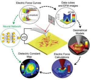
In this new method, researchers used a machine learning algorithm which once trained, was able to generate a nanoscale biochemical composition map of eukaryotic cells, from electric images obtained by high-powered scanning force microscopy. The type of algorithm used, called neural networks, mimics the way that neurons in the human brain operate, and allowed to generate information about the sample without adding external substances, one of the main goals in the cell biology field.
“It is the first study to provide such a rapid nanoscale biochemical composition map from dielectric measurements of dry eukaryotic cells, which are classically seen as being extremely difficult to map due to their complex three-dimensional topography”.
Martí Checa, IBEC researcher and first author of the work.
You can read the full article on our website here.
Press clipping:


