 ibecbarcelona.eu
- ABOUT
- STAFF
- PROJECTS
- PUBLICATIONS
- EQUIPMENT
- COLLABORATIONS
- NEWS
- JOBS
ABOUT
The main goal of the Nanoscale Bioelectrical Characterization group is to develop a multiscale and multimodal (electrical, mechanical) approach to Bioelectricity, covering from the nano- to the microscale. To this end the group combines methods and techniques from Scanning Probe Microscopy, Artificial Intelligence and Organic Bioelectronics. The main objective is to contribute to develop new label-free characterization tools for Life Sciences, new nanomedical diagnosis approaches and new electronic biosensors.
Autonomous multimodal scanning probe microscopes for Life Sciences
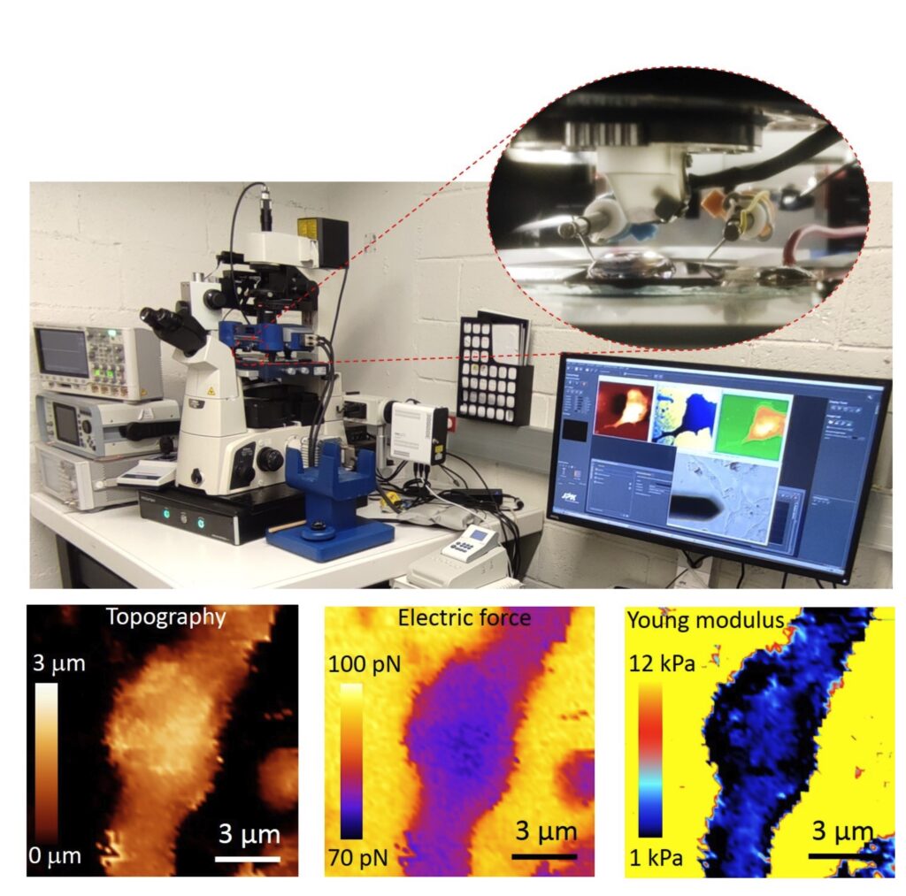
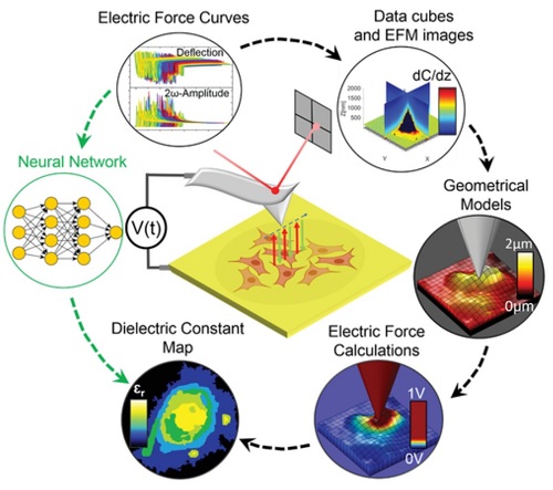
At present the group focuses in the development of an Autonomous Multimodal Functional Scanning Probe Microscope assisted by Artificial Intelligence for Life Sciences and Medical applications. The objective is to map the structural, electrical and mechanical properties at the nanoscale of cells, bacteria, drug nanocarriers and organic Bioelectronic devices with minimal intervention of the operator and at high throughput.
The objective is to obtain in an autonomous way fast functional electric and mechanical nanoscale maps of Life Science samples and Organic Electronics devices in physiological conditions with minimal intervention of the operator and at high throughput.
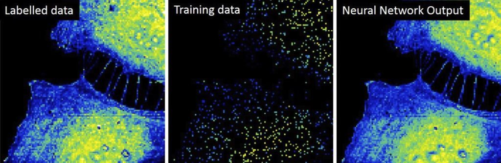
Initial results obtained by the group include the upgrade of the Scanning Dielectric Microscope to enable its operation in physiological buffers for living cell imaging, the development of a supervised machine learning algorithm to process Scanning Dielectric Microscopy data and provide almost instantaneously local dielectric constant maps of both eukaryotic and prokaryotic cells, and the implementation of a workflow for Scanning Dielectric Microscopy for high throughput and automatic nanoscale multimodal (electrical and mechanical) characterization.
High throughput multimodal characterization of drug nanocarriers
The development of novel drug nanocarriers require an exhaustive multiparametric characterization, which includes its morphology and structure, net charge, particle size distribution or phase transition temperature. These characteristics are obtained usually from different techniques. We target to obtain simultaneously and at high throughput multiparametric information on drug nanocarriers by using a single instrument, namely, the autonomous multimodal in liquid Scanning Dielectric Microscope. We aim at obtaining information on the size, sphericity, membrane wall thickness, lamellarity, Young’s modulus, stiffness, surface charge and membrane specific capacitance of drug nanocarriers, such as liposomes, polymeric nanoparticles or lipid nanoparticles.
Interrelation of mechanical and electrical processes in living neurons
Mechanical and electrical processes in cells and tissues can sometimes appear interrelated, as for instance, in the action potential propagation in neurons, which provokes the electrical polarization of the cell membrane and, at the same time, a change in neuron’s membrane tension. Similarly, the restructuring of the cytoskeleton of neurons, as occurring in the Alzheimer disease, can induce a change in cellular stiffness and, consequently, an improper neuron firing. We aim at investigating this interrelation by means of the multimodal in liquid Scanning Dielectric Microscope applied to living neurons.
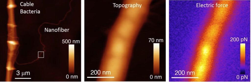
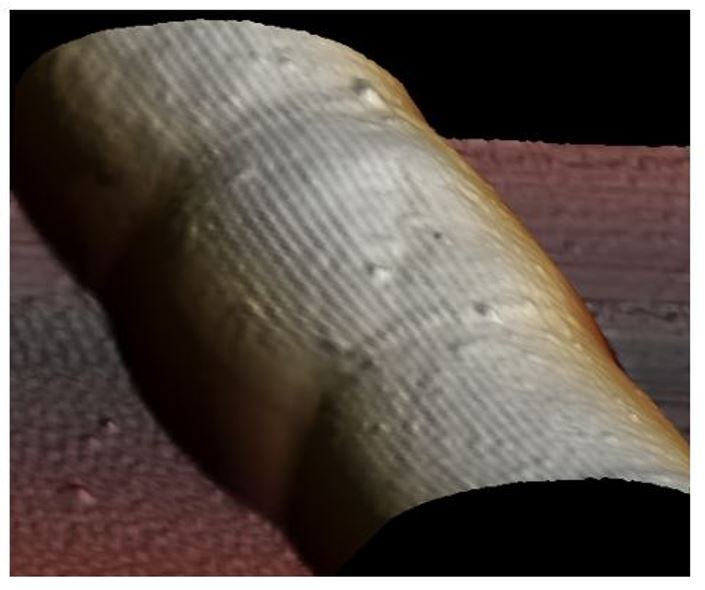
Unravelling the electrical conduction properties of cable bacteria
Long-range electron conduction in cable bacteria filaments presents unusual characteristics in the biological world, exceeding by more than 6 orders of magnitude the conductivity of the best conducting protein nanowires. Electric conduction takes place through Niquel rich protein nanofibers located in the bacteria periplasm, but still many aspects of the electronic conduction in cable bacteria remain unknown. We aim at providing new insights on the conducting properties of cable bacteria by using the unique capabilities and versatility of the Scanning Dielectric Microscope.
Novel nanoscale physical phenotyping of cancer cells
The whole process of cancer aggression, from local growth to extravasation into blood vessels, migration, seeding into different organs and formation of metastases involves physical changes (mechanical and electrical) and their interplay with protein expression and genetic transformations. We aim at developing a high throughput nanoscale multimodal physical phenotyping method for cancer cells based on the Scanning Dielectric Microscope. Our ling term objective is to provide additional diagnostics tools to medical doctors for evaluating cancer progression and aggression.
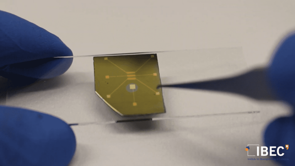
Structure-function relationships for materials in Organic Bioelectronics
Organic semiconductor materials have emerged as key materials in the development of platforms (e.g. electrolyte gated transistors) for transducing and amplifying biological and biochemical signals. This fact makes them an integral part of diverse biosensing and bioelectronic devices able to sense even single molecules or to record bioelectric potentials from excitable cells. The fundamental understanding of the nanoscale electronic and ionic transport governing the operation of these materials and devices remains, however, poorly understood. We aim at providing new insights into the structure-function relationship of organic materials used in Bioelectronics with the unique capabilities of the multimodal in operando in-liquid Scanning Dielectric Microscope.