
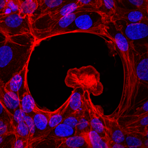
Binucleated cells could be the key in heart regeneration
A research team led by the IBEC, in collaboration with the CMR [B], discovers a mechanism that generates binucleated cells.This mechanism has been identified during the regeneration of the heart of the zebrafish, and could be associated with the extraordinary regenerative power of this animal.
After an acute heart lesion, such as a myocardial infarction, the human heart is unable to regenerate. The adult cardiac cells cannot grow and divide to replace the damaged ones, and the lesion becomes irreversible. But this does not happen in all animals. A freshwater fish native to Southeast Asia, known as a zebrafish, can completely regenerate its heart even after 20% ventricular amputation.

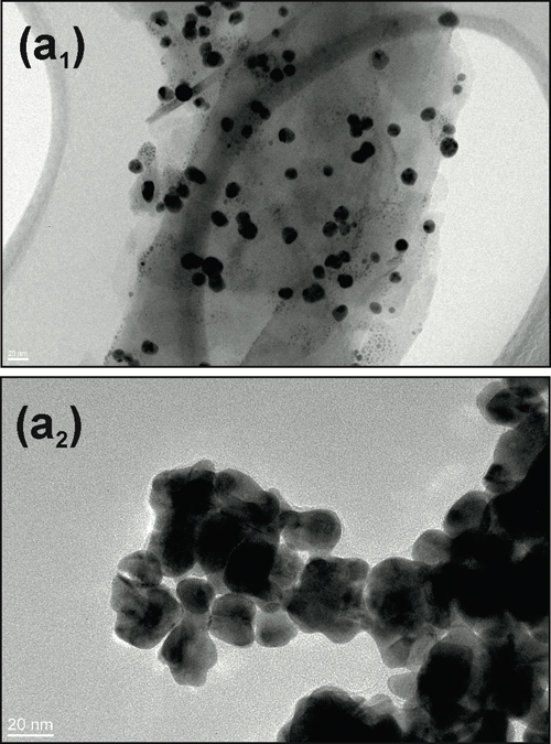 The Bacterial Infections: Antimicrobial Therapies group from IBEC, led by Eduard Torrents, has designed a new method that, for the first time, makes it possible to check antimicrobial treatment efficacy in the presence of nanoparticles.This new technique has recently been published in the Journal of Nanobiotechnology..
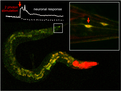 A scientific team led by IBEC and UAB manages to efficiently activate molecules located inside cell tissues using two-photon excitation of with infrared light lasers. The results of the study has been published in Nature Communications.
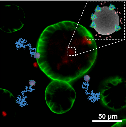
 IBEC’s
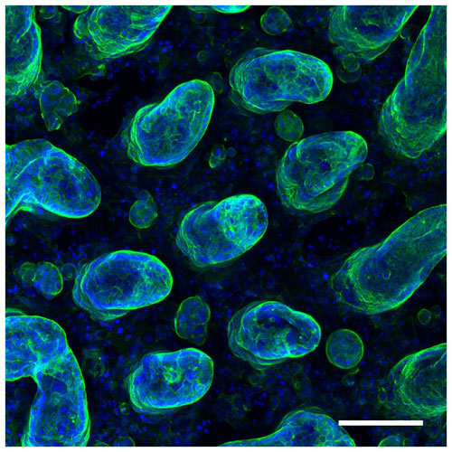
 Researchers from the IBEC have created, for the first time, 3D organoid cultures from pluripotent stem cells, which resemble human embryonic kidney tissue during the second trimester of pregnancy.
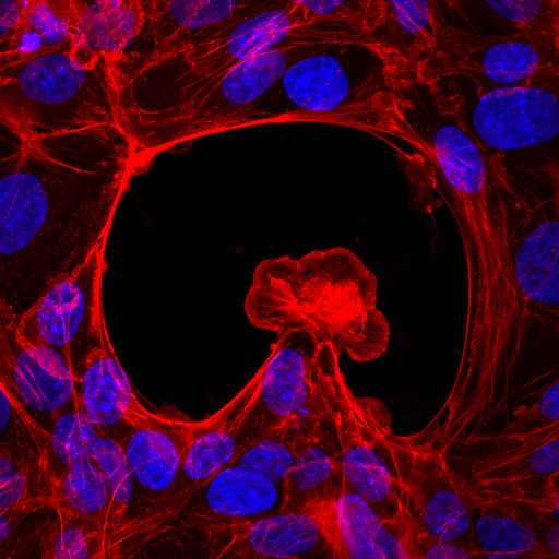
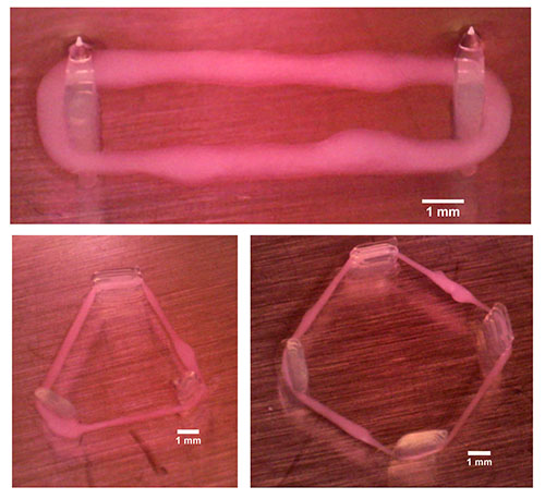
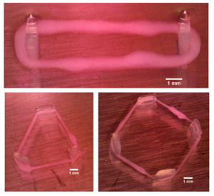 IBEC’s Smart Nano-Bio-Devices group – the institute’s experts in micro- and nanorobots – have used 3D bioprinting to produce ‘biorobots’ made of biological elements such as muscle tissue.